Note: Descriptions are shown in the official language in which they were submitted.
CA 02213861 1997-08-26
WO 96/26715 PCT/CA96/00120
~ METHOD FOR LOADING LIPID VESICLES
FIELD OF THE INVENTION
This invention relates to a method for the preparation of Iiposome
formulations of pharmaceutical compounds, and in particular to the liposome
formulations incorporating compounds bearing an amino group. This method
avoids
conditions and procedures which are detrimental to the lipids and/or
therapeutic agents or
substances to be encapsulated therein. More importantly, the present invention
provides
methods having broader applicability than has been demonstrated or described
for
previously known methods.
BACKGROUND OF THE INVENTION
Liposomes, or lipid vesicles, are a recognized drug delivery system which
can improve the therapeutic activity and increase the safety of a number of
different
pharmaceutical agents. To be useful in medical treatments, liposome
formulations should
have an efficient drug to lipid ratio, a practical shelf life and be capable
of reproducible
preparation. High drug to lipid ratios reduce the non-therapeutic lipid "load"
to the
patient, and also lowers the cost of manufacture, since less pharmaceutical is
lost in the
process of manufacture.
Liposomal carrier systems (vesicles) are microscopic spheres of one or
more lipid bilayers arranged concentrically around an aqueous core. The
vesicles have
been shown to be suitable as carriers for both hydrophilic and hydrophobic
therapeutic
agents owing to their unique combination of lipophilic and hydrophilic
portions. The
structure of the lipid bilayer is similar to the membranes enveloping animal
cells, and are
a result of amphipathic lipids arranged such that the hydrophobic portions of
the lipid
orient 'toward the center of the bilayer while the hydrophilic headgroups
orient towards
the inner or outer aqueous phases.
CA 02213861 1999-10-06
2
Liposome formulations for pharmaceutical applications can be made either
by combining drug and lipid before formation of the vesicles, or by 'loading"
lipid
vesicles with drug after they are formed. Upon administration to a patient,
liposomes
biodistribute and interact with cells in the body according to route of
administration,
vesicular composition, and vesicular size. Charge, chemistry, and the
inclusion on the
vesicle surface of protective polymers or targeting moieties, all change the
way
liposomes behave in the patient.
Despite the earlier pioneering research in developing liposome
formulations for pharmaceutical use, the further development of liposomes to
administer
pharmaceuticals has presented problems with regard to both drug encapsulation
in the
manufacturing process and drug release from the vesicle during therapy.
For drug encapsulation, there is a need to increase the trapping efficiency
such that the drug to lipid ratio is as high as possible, while maintaining
the original
chemical integrity of both drug and lipid. Consequently, the drug loading
process should
be mild and not subject the lipids, liposomes or drugs to harsh conditions
such as
extreme pH, high temperatures, or both. Once administration to a patient has
occurred,
drug release is a factor. Rapid release of pharmaceuticals from liposomes
reduces the
biodistribution benefits sought in utilizing lipid vesicle carriers.
Accordingly, efforts to
optimize pharmaceutical loading and to reduce the rate of release of
pharmaceuticals
from lipid vesicles have continued. For clinical applications, the liposome
formulations
should also be capable of existing stably in a formulated state or in a ready-
to-mix ldt to
allow for shipping and storage.
What is needed in the art are new methods for the preparation of stable
liposome formulations of therapeutic agents which are easy to prepare, provide
suitable
retention of the therapeutic agent, and which provide high drug to lipid
ratios. Quite
surprisingly, the present invention fulfills these and other needs.
CA 02213861 1999-10-06
2a
SUMMARY OF INVENTION
This invention provides a method of preparing a liposome formulation of a
protonatable therapeutic agent, said method comprising:
(i) preparing a. mixture of liposomes in an aqueous solution, said liposomes
having an encapsulated medium and an external medium, wherein said
encapsulated medium
and said external medium each contain a methylammonium salt;
(ii) establishing a concentration gradient of methylamine across the liposome
membranes by removing or diluting said methylammonium salts in said external
medium; and
1 o (iii) incubating said liposomes of step (ii) with said protonatable
therapeutic agent,
said protonatable therapeutic agent being present in a neutral form which is
attracted toward
said encapsulated medium of said liposomes by said concentration gradient of
methylamine,
for a period of time sufficient to cause adherence of said therapeutic agent
to said liposomes.
This invention also provides the use of liposome formulations prepared
according to
the preceding method for treatment, such as treatment of neoplasms and
micobacterial
infections.
This invention also provides liposomal formulations prepared according to the
preceding method.
This invention also provides a method of retaining a protonatable therapeutic
agent in
2 0 a liposome formulation, said method comprising:
(i) preparing a mixture of liposomes in an aqueous solution, said liposomes
having an encapsulated medium and an external medium, wherein said
encapsulated medium
and said external medium contain a methylammonium salt and a therapeutic
agent; and
(ii) establishing a concentration gradient of methylamine across the liposome
2 5 membranes by removing or diluting said methylammonium salt from said
external medium,
wherein said gradient results in said encapsulated therapeutic agents becoming
protonated and
thereby retained in said liposome formation.
The present invention provides methods for the preparation of stable liposome
formulations of protonatable therapeutic agents. The method involves loading a
therapeutic
3 o agent into performed liposomes having a methylamine concentration gradient
CA 02213861 1997-08-26
WO 96126715 PCT/CA96/00120
3
across the lipid bilayer of the liposomes. This method provides liposome
formulations
which are more stable, more cost effective, and easier to prepare in a
clinical
environment than those previously available. Additionally, these methods have
application to a broader spectrum of pharmaceutical agents than methods
previously
. 5 described. The present invention also provides the pharmaceutical
compositions prepared
by the above method, a kit for the preparation of liposome formulations of
therapeutic
agents, and methods for their use.
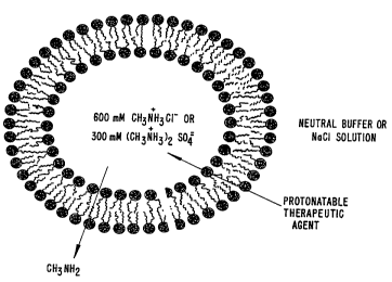
Although not intending to be bound by any particular theory, it is
postulated that neutral therapeutic agents can be loaded into preformed
liposomes having
a methylamine concentration gradient across their lipid bilayer by a mechanism
illustrated
in Figure 1. In this particular embodiment, liposomes are formed having an
encapsulated
medium which contains a methylammonium salt. The external medium which
originally
has the composition of the encapsulated medium is exchanged with a neutral
external
medium. A therapeutic agent such as doxorubicin or ciprofloxacin (structures
shown in
Figure 2) which is both lipophilic and which can be protonated is drawn toward
the
liposomes' encapsulated medium by both its polarity and the methylammonium ion
gradient which is established across the bilayer. Methylamine diffuses out of
the
liposomes as the therapeutic agents are drawn in and protonated, thereby
maintaining the
differential (see Figure I).
The liposomes of this invention may be prepared with therapeutic agents
which precipitate at pH ranges usual in the preparation and loading of lipid
vesicles by
converational means. Further advantages for these liposomal compositions
include a long
shelf life which is a result of the lipids not being exposed to harsh
conditions which can
hydrolyze them.
The present invention also provides a method for reducing the rate of
release of a protonatable therapeutic agent from Iipid vesicles. In this
method, a
transmembrane potential, oriented to retain the agent in the lipid vesicles,
is generated
across the lipid vesicle membranes. As described in detail below, it has been
surprisingly found that such a methylamine gradient is capable of producing
over a
thousand-fold reduction in the rate of release of protonatable therapeutic
agents such as
ciprofloxacin and doxorubicin from liposomes. Additionally, the method can be
used
with essentially any protonatable material which can be encapsulated in a
lipid vesicle.
The methylamine gradient can be generated after encapsulation of the
protonatable
CA 02213861 1997-08-26
WO 96126715 PCT/CA96/00120
4
compound or can be the result of lipid vesicle formation.
In addition to the foregoing methods, the invention also provides the
products produced by practicing the methods, namely pharmaceutical
preparations
comprising a pharmaceutical agent which has been loaded into lipid vesicles by
means of
a methylamine gradient.
The present invention provides fast, safe, stable, efficient and inexpensive
methods for loading amphipathic therapeutic agents into liposomes. The
resulting
compositions release the therapeutic agent slowly in the patient and therefore
achieve
maximal efficacy in vivo. The bitter taste and smell of pharmaceuticals such
as
ciprofloxacin can be masked using the invention, and it can be used to load
drugs that
are hydrochloride salts and therefore have a low solution pH. As a result, the
present
invention can be used to prepare pharmaceutical compositions that can not be
generated
in large quantities by any other technique.
BRIEF DESCRIPTION OF THE DRAWINGS
Figure 1 shows a model of a liposome used to load a protonatable
therapeutic agent.
Figure 2 shows the structures of doxorubicin and ciprofloxacin, two
therapeutic agents which can be Ioaded and retained in liposomes according to
the present
invention.
Figure 3 shows the temperature dependent uptake and retention of
doxorubicin in LUVs exhibiting 600 mM ethanolamine HCl gradients.
Figure 4 shows the temperature dependent uptake and retention of
vincristine in LUVs exhibiting ethanolamine HCl gradients.
Figure 5 shows the temperature dependent uptake and retention of
cipro~oxacin in LUVs exhibiting 600 mM ethanoIamine HCl gradients.
Figure 6 shows the uptake and retention of doxorubicin, epirubicin, and
vincristine in LUVs (DPPC/chol) exhibiting a 600 mM EDAS gradient at
60°C.
Figure 7 shows the uptake of ciprofloxacin in POPC/chol LUVs exhibiting ,
a 300 mM EDAS gradient with an external pH 5.5 citrate buffer.
Figure 8 shows the uptake of ciprofloxacin in DPPC/chol LUVs in
response to amine gradients at 60°C.
CA 02213861 1997-08-26
WO 96!26715 PCT/CA96/00120
Figure 9 shows the temperature dependent uptake of tryptophan in
response to 300 mM methylammonium sulfate gradients.
Figure l0A shows the uptake of doxorubicin in 100 nm size unilamellar
vesicles (LUVs) composed of egg phosphatidylcholine and formed in the presence
of a
5 gradient of 300 mM CH3NH3Cl (methylammonium chloride or MAC), at a drug to
lipid
(molar) ratio of 0.26 and a temperature of 25°C.
Figure lOB show the improved retention of doxorubicin in egg
phosphatidylcholine LUVs under the same conditions as in Figure l0A except the
liposomes were formed in MAC at a concentration of 600 mM.
Figure lOC shows drug retention characteristics of the loaded formulation
from Figure lOB over the 24 hours following loading.
Figure 11 A depicts the uptake observed for doxorubicin at a drug to lipid
ratio of 0.13 over approximately 30 minutes at 600 mM of MAC in
DPPC/cholesterol
vesicles at 45 ° C.
Figure 11B is a graph showing drug retention over a period of 24 hours
for the loaded formulation of Figure 11A.
Figure 12A illustrates the loading of doxorubicin obtained with 200 nm
DPPC/cholesterol LUVs prepared in 600 mM MAC. The drug to lipid ratio was
initially
0.25, and the temperature was 45°C.
Figure 12B is a graph showing the drug retention characteristics of the
formulations of Figure 12A.
Figure 13 shows the loading characteristics of doxorubicin into
sphingomyelin/ceramide/cholesterol 4:1:4 (molar ration of Sph/Cer/cholesterol)
LUVs of
200 nm prepared in 600 mM MAC. The temperature was 45°C and the drug to
lipid
ratio was 0.36.
Figure 14 illustrates the uptake of ciprofloxacin into 200 nm
DPPC/cholesterol(55:45, mol:mol) LUVs in response to an internal concentration
of 600
mM MAC at 45°C for an initial drug to lipid (molar) ratio of 2.9.
Figure 15 shows the results of loading of ciprofloxacin into
distearoylphosphatidylcholine/cholesterol 55:45 mol: mol (DSPC)/cholesterol
100 nm
LUVs in response to a 300 mM (CH3NH3)ZS04 (MAS) gradient at 65°C and
with a drug
to lipid ratio of 0.5. The final drug to lipid ratio was 0.4.
CA 02213861 1997-08-26
WO 96/26715 PCT/CA96/00120
6
Figure 16 illustrates the uptake of doxorubicin in DPPC/cholesterol (55:45
mol:mol) into 100 nm LUVs prepared in 300 mM MAS at 45°C, with initial
drug to
lipid ratios of 0.37 (Figure 16A) and 1.2 (Figure 16B).
Figure 17 is a graph showing the uptake of doxorubicin into '
Sph/C'.er/cholesterol 1:1:1 (molar ratio) 100 nm LUVs containing Peg 2000Cer-
C14
prepared in 300 mM MAC at 50°C, with an initial drug to lipid ratio of
0.66.
Figure 18 shows the retention characteristics exhibited by liposomes loaded
with doxorubicin into DSPC/Cholesterol + 2 % Peg2000Cer-C20 + 1
N-(4-p-maleimidophenyl)butyryl)phosphatidylethanolamine (MPB-PE) lipid
vesicles using
a 300 mM methylammonium sulfate gradient. The three curves show lipid vesicles
(circles), lipid vesicles preincubated in 50% mouse serum (squares), and
antibody-coupled lipid vesicles incubated 50 % mouse serum (triangles).
DETAILED DESCRIPTION OF THE INVENTION
CONTENTS
I. Glossary
II. General
III. Methods of Loading Therapeutic Agents Into Liposomes
IV. Methods of Retaining Therapeutic Agents In Liposomes
V. Pharmaceutical Formulations
VI. Administration of Liposome Formulations
VII. Examples
VIII. Conclusion
I. Glossary
The following abbreviations are used herein: DOX, doxorubicin; Cer,
ceramide; Chol, cholesterol; CIP, ciprofloxacin; DPPC, dipalmitoyl
phosphatidylcholine;
DSPC, distearoyl phosphatidylcholine; DTT, dithiothreitol; EDAS,
ethylenediammonium
sulfate; HBS, Hepes buffered saline; LUV, large unilamellar vesicles; MAC,
CA 02213861 1997-08-26
WO 96126715 PCTlCA96/00120
7
methylammonium chloride; MAS, methylammonium sulfate; MLV, multilamellar
vesicles; MPB, 4-(maleimidophenyl)butyryl; PE, phosphatidylethanolamine; PEG,
polyethylene glycol; PEG-Cer-CZO, 1-O-(2'-(w-
methoxypolyethyleneglycol)succinoyl)-2-
N-arachidoyl-sphingosine; PEG-Cer-C,4, 1-O-(2'-(w-methoxypolyethylene-
glycol)succinoyl)-2-N-myristoyl-sphingosine; POPC,
palmitoyloleoylphosphatidylcholine;
and Sph, sphingomyelin.
As used herein, the terms "pharmaceutical preparation" and "protonatable
agent," have the following meanings: pharmaceutical preparation means a
composition of
matter suitable for administration to humans or animals and comprising a
biologically
active material and appropriate buffers, diluents, carriers, and the like; and
protonatable
agent means an agent which can exist in a charged state when dissolved in an
aqueous
medium. Examples of protonatable agents include compounds having an amino
group
which can be protonated in acidic media, and compounds which are zwitterionic
in
neutral media (i. e. , amino acids and agents such as ciprofloxacin) and which
can also be
protonated in acidic environments.
The term "lipid" refers to any suitable material resulting in a bilayer such
that a hydrophobic portion of the lipid material orients toward the bilayer
while a
hydrophilic portion orients toward the aqueous phase. Amphipathic lipids are
necessary
as the primary lipid vesicle structural element. Hydrophilic characteristics
derive from
the presence of phosphato, carboxylic, sulfato, amino, sulfhydryl, nitro, and
other like
groups. Hydrophobicity could be conferred by the inclusion of groups that
include, but
are not limited to, long chain saturated and unsaturated aliphatic hydrocarbon
groups and
such groups substituted by one or more aromatic, cycloaliphatic or
heterocyclic group(s).
The preferred amphipathic compounds are phosphoglycerides and sphingolipids,
representative examples of which include phosphatidylcholine,
phosphatidylethanolamine,
phosphatidylserine, phosphatidylinositol, phosphatidic acid, palmitoyloleoyl
phosphatidylcholine, lysophosphatidylcholine, lysophosphatidylethanolamine,
dipalmitoylphosphatidylcholine, dioleoylphosphatidylcholine,
distearoylphasphatidylcholine or dilinoleoylphosphatidylcholine could be used.
Other
compounds lacking in phosphorus, such as sphingolipid and glycosphingolipid
families
are also within the group designated as lipid. Additionally, the ampipathic
lipids
described above may be mixed with other lipids including triglycerides and
sterols.
CA 02213861 1997-08-26
WO 96126715 PCT/CA96/00120
8
As used herein, the term "tissue" refers to any pathological cell or group
of cells that exhibit a similar pathological trait. For example, the trait may
be malignant
transformation. Alternatively, the trait may be intracellular infection.
Generally, the
tissue will include cells that originate from the same cell type.
II. General
The liposomes which are used in the present invention are formed from
standard vesicle-forming lipids, which generally include neutral and
negatively charged
phospholipids and a sterol, such as cholesterol. The selection of lipids is
generally
guided by consideration of, e. g. , liposome size and stability of the
liposomes in the
bloodstream.
Typically, the major lipid component in the liposomes is
phosphatidylcholine. Phosphatidylcholines having a variety of acyl chain
groups of
varying chain length and degree of saturation are available or may be isolated
or
synthesized by well-known techniques. In general, less saturated
phosphatidylcholines
are more easily sized, particularly when the liposomes must be sized below
about 0.3
microns, for purposes of filter sterilization. Phosphatidylcholines containing
saturated
fatty acids with carbon chain lengths in the range of C,4 to C2z are
preferred.
Phosphatidylcholines with mono or diunsaturated fatty acids and mixtures of
saturated
and unsaturated fatty acids may also be used. Other suitable lipids include
phosphonolipids in which the fatty acids are linked to glycerol via ether
linkages rather
than ester linkages. Liposomes useful in the present invention may also be
composed of
sphingomyelin or phospholipids with head groups other than choline, such as
ethanolamine, serine, glycerol and inositol. Preferred liposomes will include
a sterol,
preferably cholesterol, at molar ratios of from 0.1 to 1.0
(cholesterol:phospholipid).
Most preferred Iiposome compositions are
distearoylphosphatidylcholine/cholesterol,
dipalmitoylphosphatidylcholine/cholesterol, and sphingomyelin/cholesterol.
Methods
used in sizing and filter-sterilizing liposomes are discussed below.
The liposomes can be prepared by any of the techniques now known or
subsequently developed for preparing liposomes. For example, the liposomes can
be
formed by the conventional technique for preparing multilamellar lipid
vesicles (MI,Vs),
CA 02213861 1999-10-06
9
that is, by depositing one or more selected lipids on the inside walls of a
suitable vessel
by dissolving the lipids in chloroform and then evaporating the chloroform,
and by then
adding the aqueous solution which is to be encapsulated to the vessel,
allowing the
aqueous solution to hydrate the lipid, and swirling or vortexing the resulting
lipid
suspension. This process engenders a mixture including the desired liposomes.
Alternatively, techniques used for producing large unilamellar lipid
vesicles (L,UVs), such as reverse-phase evaporation, infusion procedures, and
detergent
dilution, can be used to produce the liposomes. A review of these and other
methods for
producing lipid vesicles c;an be found in the text Liposome Technology, Volume
I,
Gregory Gregoriadis Ed., CRC Press, Boca Raton, Florida, (1984).
For example, the lipid-containing particles can be in
the form of steroidal lipid vesicles, stable plurilamellar lipid vesicles
(SPLVs),
monophasic vesicles (MPVs), or lipid matrix carriers (LMCs) of the types
disclosed in
Lenk, et al. U.S. Patent No. 4,522,803, and Fountain, et al. U.S. Patent Nos.
4,588,578
and 4, 610, 868 . A
particularly preferred method for preparing LUVs is described in U.S. Patent
No.
5,008,050.
Additionally, in the case of MLVs, if desired, the liposomes can be
subjected to multiple (five or more) freeze-thaw cycles to enhance their
trapped volumes
and trapping efficiencies and to provide a more uniform interlamellar
distribution of
solute (Mayer, et al. , J. Biol. Chem. 260:802-808 ( 1985)).
Following liposome preparation, the liposomes may be sized to achieve a
desired size range and relatively narrow distribution of liposome sizes. A
size range of
about 0.2-0.4 microns allows the liposome suspension to be sterilized by
filtration
through a conventional filter, typically a 0.22 micron filter. The filter
sterilization
method can be carried out on a high through-put basis if the liposomes have
been sized
down to about 0.2-0.4 microns.
Several techniques are available for sizing liposomes to a desired size.
One sizing method is described in U.S. Pat. No. 4,737,323.
Sonicating a liposome suspension either by bath or probe sonication produces
a progressive size reduction down to small unilamellar vesicles less than
about 0.05
microns in size. Homogenization is another method which relies on shearing
energy to
fragment large liposomes into smaller ones. In a typical homogenization
procedure,
CA 02213861 1999-10-06
multilamellar vesicles are recirculated through a standard emulsion
homogenizer until
selected liposome sizes, typically between about 0.1 and 0.5 microns, are
observed. In
both methods, the particle size distribution can be monitored by conventional
laser-beam
particle size determination.
5 Extrusion of liposome through a small-pore polycarbonate membrane or an
asymmetric ceramic membrane is also an effective method for reducing liposome
sizes to
a relatively well-defined size distribution (see, U.S. Patent No. 5,008,050
and Hope, et
al., in: Liposome Technology, vol. I, 2d ed. (G. Gregoriadis, Ed.) CRC Press,
pp.
123-139 ( 1992) ,
10 Typically, the suspension is cycled through the membrane one or more times
until the
desired liposome size distribution is achieved. The liposomes may be extruded
through
successively smaller-pore membranes, to achieve a gradual reduction in
liposome size.
For use in the present inventions, liposomes having a size of from about 0.05
microns to
about 0.15 microns are preferred. Other useful sizing methods such as
sonication,
solvent vaporization or reverse phase evaporation are known to those of skill
in the art.
Liposomes prepared in the method of the invention may be dehydrated for
longer storage. For this purpose, two basic approaches are provided. In one
approach,
the lipid vesicles are loaded with the therapeutic agent according to the
method of the
invention, dehydrated for purposes of storage, shipping, and the like, and
then
rehydrated at the time of use. Alternatively, the lipid vesicles may be formed
without
the protonatable therapeutic agent, dehydrated for storage, as above, and then
at or near
the time of use, rehydrated using a solution of the protonatable therapeutic
agent such
that the methylamine gradient is created, and the pharmaceutical agent is
loaded.
In either case, the liposomes are preferably dehydrated under reduced
pressure using standard freeze-drying equipment or equivalent apparatus. The
lipid
vesicles and their surrounding medium can also be frozen in liquid nitrogen
before being
dehydrated or not, and placed under reduced pressure. Dehydration without
prior
freezing takes longer than dehydration with prior freezing, but the overall
process is
gentler without the freezing step, and thus there is subsequently less damage
to the lipid
vesicles and a smaller loss of the internal contents. Dehydration without
prior freezing at
room temperature and at a reduced pressure provided by a vacuum pump capable
of
producing a pressure of about 1 mm Hg typically takes between approximately 24
and 36
hours, while dehydration with prior freezing under the same conditions
generally takes
CA 02213861 1999-10-06
11
between approximately 12 and 24 hours.
To ensure that the liposomes will survive the dehydration process without
losing a substantial portion of their internal contents, it is important that
one or more
protective sugars be available to interact with the lipid vesicle membranes
and keep them
intact as the water in the system is removed. A variety of sugars can be used,
including
such sugars as trehalose, maltose, sucrose, glucose, lactose, and dextran. In
general,
disaccharide sugars have been found to work better than monosaccharide sugars,
with the
disaccharide sugars trehalose and sucrose being most effective. Other more
complicated
sugars can also be used. For example, aminoglycosides, including streptomycin
and
dihydrostreptomycin, have been found to protect lipid vesicles during
dehydration.
Typically, one or more sugars are included as part of either the internal or
external media of the lipid vesicles. Most preferably, the sugars are included
in both the
internal and external media so that they can interact with both the inside and
outside
surfaces of the liposomes' membranes. Inclusion in the internal medium is
accomplished
by adding the sugar or sugars to the buffer which becomes encapsulated in the
lipid
vesicles during the lipid vesicle formation process. Since in most cases this
buffer also
forms the bathing medium for the finished lipid vesicles, inclusion of the
sugars in the
buffer also makes them part of the external medium. Of course, if an external
medium
other than the original buffer is used, e.g., to create a transmembrane
potential (see
above), the new external medium should also include one or more of the
protective
sugars.
The amount of sugar to be used depends on the type of sugar used and the
characteristics of the lipid vesicles to be protected. See, U.S. Patent No.
4,880,635 and
Harrigan, et al. , Chem. Phys. Lipidr 52:139-149 ( 1990) ,
. Persons skilled in the art can readily test various sugar
types and concentrations to determine which combination works best for a
particular lipid
vesicle preparation. In general, sugar concentrations on the order of 100 mM
and above
have been found necessary to achieve the highest levels of protection. In
terms of moles
of membrane phospholipid, millimolar levels on the order of 100 mM correspond
to
approximately 5 moles of sugar per mole of phospholipid.
Lt the case of dehydration without prior freezing, if the lipid vesicles being
dehydrated are of the type which have multiple lipid layers and if the
dehydration is
carried to an end point where between about 2 % and about 5 ~O of the original
water in
CA 02213861 1999-10-06
12
the preparation is left in the preparation, the use of one or more protective
sugars may be
omitted.
Once the lipid vesicles have been dehydrated, they can be stored for
extended periods of time until they are to be used. The appropriate
temperature for
storage will depend on the make up of the lipid vesicles and the temperature
sensitivity
of whatever materials have been encapsulated in the lipid vesicles. For
example, as is
known in the art, various pharmaceutical agents are heat labile, and thus
dehydrated lipid
vesicles containing such. agents should be stored under refrigerated
conditions so that the
potency of the agent is not lost. Also, for such agents, the dehydration
process is
preferably carried out at reduced temperatures, rather than at room
temperature.
In certain embodiments, it is desirable to target the liposomes of the
invention using targeting moieties that are specific to a particular cell
type, tissue, and
the like. Targeting of liposomes using a variety of targeting moieties (e.g.,
ligands,
receptors and monoclonal antibodies) has been previously described (see, e.g.,
U.S.
Patent Nos. 4,957,773 and 4,603,044.
Examples of targeting moieties include monoclonal antibodies specific to
antigens associated with neoplasms, such as prostate cancer specific antigen.
Tumors can
also be diagnosed by detecting gene products resulting from the activation or
overexpression of oncogenes, such as ras or c-erB2. In addition, many tumors
express
antigens normally expressed by fetal tissue, such as the alphafetoprotein
(AFP) and
carcinoembryonic antigen (CEA). Sites of viral infection can be diagnosed
using various
viral antigens such as hepatitis B core and surface antigens (HBVc, HBVs)
hepatitis C
antigens, Epstein-Barr virus antigens, human immunodeficiency type-1 virus
(HIV1) and
papilloma virus antigens. Inflammation can be detected using molecules
specifically
recognized by surface molecules which are expressed at sites of inflammation
such as
integrins (e.g., VCAM-1), selectin receptors (e.g., ELAM-1) and the like.
Standard methods for coupling targeting agents to liposomes can be used.
These methods generally involve incorporation into liposomes of lipid
components, such
as phosphatidylethanolamine, which can be activated for attachment of
targeting agents,
or derivatized lipophilic compounds, such as lipid derivatized bleomycin.
Antibody
targeted liposomes can be constructed using, for instance, liposomes which
incorporate
protein A (see, Renneisen, et al. , J. Biol. Chem. , 265:16337-16342 (1990)
and Leonetti,
CA 02213861 1999-10-06
13
et al. , Proc. Natl. Acad. Sci. (ZISA) 87:2448-2451 ( 1990) .
Targeting mechanisms generally require that the targeting agents be
positioned on the surface of the liposome in such a manner that the target
moieties are
available for interaction with the target, for example, a cell surface
receptor. The
liposome is typically fashioned in such a way that a connector portion is
first
incorporated into the membrane at the time of forming the membrane. The
connector
portion must have a liF~ophilic portion which is firmly embedded and anchored
in the
membrane. It must also have a hydrophilic portion which is chemically
available on the
aqueous surface of the liposome. The hydrophilic portion is selected so that
it will be
chemically suitable to form a stable chemical bond with the targeting agent
which is
added later. Therefore, the connector molecule must have both a lipophilic
anchor and a
hydrophilic reactive group suitable for reacting with the target agent and
holding the
target agent in its corr~~t position, extended out from the liposome's
surface. In some
cases it is possible to attach the target agent to the connector molecule
directly, but in
most instances it is more suitable to use a third molecule to act as a
chemical bridge,
thus linking the connecaor molecule which is in the membrane with the target
agent
which is extended, three dimensionally, off of the vesicle surface.
III. Methods of Loadins Therapeutic Agents Into Liposomes
Traditional methods of loading conventional drugs into liposomes include
an encapsulation technique and a transmembrane potential loading method. In
the
encapsulation technique, the drug and liposome components are dissolved in an
organic
solvent or mixture of solvents in which all species are miscible, and then
concentrated to
a dry film. A buffer is then added to the dried film and liposomes are formed
having the
drug incorporated into the vesicle walls. In a modification of this
encapsulation
technique, the drug can be placed into a buffer and added to a dried film of
only lipid
components. In this manner, the drug will become encapsulated in the aqueous
interior
of the liposome. The buffer which is used in the formation of the liposomes
can be any
biologically compatible buffer solution of, for example, isotonic saline,
phosphate
buffered saline, or other low ionic strength buffers.
CA 02213861 1997-08-26
WO 96126715 PCT/CA96/00120
14
Encapsulation methods;- often referred to a "passive loading" suffer from
several limitations. One such limitation is the low drug-to-lipid (D/L) ratio
which is
achieved. With low D/L ratios, larger amounts of lipids must be administered
in order
to provide a suitable drug concentration in vivo. Related to this low D/L
ratio is the
inefficiency of drug uptake into the Iiposomes which can result in substantial
waste of the
expensive pharmaceutical agents. Alternatively, recycling and reisolation of
the
unencapsulated pharmaceutical agents results in additional costs for
preparation.
Alternative loading processes, termed "active loading," involve the use of
transmembrane potentials. Transmembrane potential loading has been described
in detail
in U.S. Patent No. 4,885,172, U.S. Patent No. 5,059,421, U.S. Patent No.
5,171,578,
U.S. Patent No. 5,316,771 and U.S. Patent No. 5,380,531. Briefly, the
transmembrane
potential loading method is used for a number of conventional drugs which can
exist in a
charged state when dissolved in an appropriate aqueous medium. Preferably, the
drug
will be relatively lipophilic so that it will partition into the liposome
membranes. The
loading process is carried out by creating a transmembrane potential across
the bilayers
of the liposomes. The transmembrane potential is generated by creating a
concentration
gradient for one or more charged species (e. g. , Na+, K+, H+ and/or NH4+)
across the
membranes. This concentration gradient is generated by producing liposomes
having
different internal and external media. Thus, for a drug which is negatively
charged when
ionized, a transmembrane potential is created across the membranes which has
an inside
potential which 'is positive relative to the outside potential. For a drug
which is
positively charged, the opposite transmembrane potential would be used.
Despite this more "active" loading process, some of these methods still
suffer from inefficient drug loading or limitations resulting from the
particular media
used. For example, Mayer, et al., J. Biol. Chem. 260:802-808 (1985), describe
the
loading of a local anesthetic dicubaine, into liposomes using Na+ and K+
gradients.
However, only 52 % loading was achieved. Methods which involve H+ ion
gradients (or
pH gradients) have proven useful for a number of liposome loading
applications.
Nevertheless, these methods also have their limitations. For example, one
method which
uses a pH gradient involves preparing liposomes having an acidic interior
medium and a
neutral exterior medium. The use of an unphysiologically acidic pH can degrade
some
drugs and also promote lipid hydrolysis and subsequent leakage of any
encapsulated
drug. Additionally, the method does not appear to be useful for those drugs
which have
CA 02213861 1997-08-26
WO 96126715 PCT/CA96/00120
both a basic amine functionality and a carboxylic acid functionality (e.g.,
amino acids,
small peptides and zwitterionic drugs). For example, Chakrabarti, et al. U.S.
Patent No
5,380,531 describes pH loading methods for amino acids and peptides in which
the
amino acids and peptides are first derivatized to their ester or amide forms.
Chakrabarti,
5 et al. also note that the method does not work for the more basic amino acid
esters and
peptide esters such as histidine methyl ester, (Lys)5 methyl ester and Lys-
(Ala)4 methyl
ester. Other problems which exist for pH loading methods involve the limited
solubility
of some drugs in a neutral external medium. For example, the quinolone
antibacterial
agent ciprofloxacin is essentially insoluble in water in the pH range 6 to 8.
If the
10 external pH is lowered to about 5 (a point at which ciprofloxacin is
adequately soluble)
the gradient is insufficient for rapidly and efficiently loading the drug.
More recently, Barenholz, et al. U.S. Patent Nos. 5,316,771 and
5,192,549 have described an active loading method which utilizes a NH4+ ion
gradient.
Barenholz, et al. note that liposome loading using a NH4+ ion gradient is
useful for weak
15 amphipathic compounds having either weak basic or acidic moieties.
Additionally,
Barenholz, et al. note that within a group of weak bases, the more hydrophobic
of the
group will load more readily. While this method provides an alternative
strategy for
loading of certain compounds, there still exists a need in the art for methods
of broader
application, for example, methods which are suitable for compounds which
possess both
basic and acidic functionality and which are relatively insoluble in water at
neutral pH.
Surprisingly, we have found that the use of methylammonium ion gradients
enable the rapid loading and retention of drugs such as ciprofloxacin into
liposomes.
Moreover, methods which use methylammonium ion gradients provide a broader
range
of loading possibilities than are available with gradients using other amines
such as
ethanolamine or glucosamine.
Accordingly, the present invention provides in one aspect a method of
preparing a stable liposome formulation of a protonatable therapeutic agent.
In this
method, liposomes are first prepared which encapsulate an aqueous solution of
a
methylammonium salt. Following preparation of the liposomes, a concentration
gradient
of methylamine across the liposome membranes is established, and the resulting
liposome
mixture is incubated with a neutral form of the protonatable therapeutic went.
The
therapeutic agent is drawn into the liposome as a result of the concentration
gradient of
methylamine, and once encapsulated, is protonated and trapped.
CA 02213861 1999-10-06
16
Liposomes which encapsulate an aqueous solution of a methylamine salt
can be prepared by any of the methods described above. Subsequent loading of
the
protonatable therapeutic, agent into the liposomes will be dependent on the
methylamine
concentration gradient (or methylammonium ion gradient) and the pH gradient
which also
results from a change in methylamine concentrations between the lipid
bilayers. The
gradients are created by forming liposomes in a methylammonium salt solution,
followed
by removal or dilution ~of the salt from the external aqueous phase of the
liposomes. A
number of methylammanium salts are useful in the present invention, including
methylammonium chloride, methylammonium sulfate, methylammonium citrate and
methylammonium acetate. Other salts which are suitable in pharmaceutical
formulations
are known to those of skill in the art. The concentration of the
methylammonium salt
solution which is encapsulated can vary from about 50 mM to about 1 M, however
concentrations of 200 mM to 800 mM are preferred, with 300 mM to 600 mM being
particularly preferred. In general an initial methylammonium ion concentration
of about
600 mM is the most preferred. To create the concentration gradient, the
original
external medium is replaced by a new external medium having a different
concentration
of the charged species or a totally different charged species. The replacement
of the
external medium can be accomplished by various techniques, such as, by passing
the
lipid vesicle preparation through a gel filtration column, e.g., a SephadeX
column, which
has been equilibrated with the new medium, or by centrifugation, dialysis, or
related
techniques.
Depending upon the permeability of the lipid vesicle membranes, the full
transmembrane potential corresponding to the concentration gradient will
either form
spontaneously or a permeability enhancing agent, e.g., a proton ionophore may
have to
be added to the bathing medium. If desired, the permeability enhancing agent
can be
removed from the preparation after loading has been completed using
chromatography or
other techniques. In either case, a transmembrane potential having a magnitude
defined
by the Nernst equation will appear across the lipid vesicles' membranes. The
change in
composition of the external phase causes an outflow of neutral methylamine
from the
interior encapsulated medium to the external medium. This outflow also results
in a
reverse pH gradient by accumulation of hydrogen ions left behind in the
internal aqueous
phase. An influx of a neutral form of a protonatable therapeutic agent into
the liposomes
replaces the methylamine.
*Trade-mark
CA 02213861 1997-08-26
WO 96/26715 PCT/CA96/00120
17
In accordance with the invention, it has also been found that the
methylamine gradient can be used to load protonatable therapeutic agents into
the lipid
vesicles regardless of the pH of the external solution. This is very
surprising in light of
' earlier loading methods. Still further, once Iipid vesicles having a
concentration gradient
S and thus a methylamine gradient of the appropriate orientation have been
prepared, the
proceas of loading pharmaceutical agents into the lipid vesicles reduces to
the very
simple step of adding the agent to the external medium. Once added, the
mei:hylamine
gradient will automatically load the agent into the lipid vesicles. As
described in detail
in thc~ Examples below, the loading is not only simple, but is also extremely
efficient.
As also described in the Examples below, it has been found that trapping
efficiencies for
pharmaceutical agents of 90 % and higher can be readily achieved with the
methylamine/methylammonium ion gradient loading technique.
The methylamine gradient loading method can be used with essentially any
therapeutic agent which can exist in a charged state when dissolved in an
appropriate
, aqueous medium (e.g., organic compounds which include an amino group which
can be
protonated and some zwitterionic compounds such as the quinolone antibacterial
agents).
Preferably, the agent, or drug, should be relatively lipophilic so that it
will partition into
the lipid vesicle membranes. Examples of some of the pharmaceutical agents
which can
be loaded into Iipid vesicles by this method include doxorubicin, mitomycin,
bleomycin,
daunorubicin, streptozocin, vinblastine, vincristine, mechlorethamine
hydrochloride,
melphalan, cyclophosphamide, triethylenethiophosphoramide, carmustine,
lomustine,
semustine, fluorouracil, hydroxyurea, thioguanine, cytarabine, floxuridine,
decarbazine,
procarbazine, and ciprofloxacin. Preferably, the drugs which are loaded into
liposomes
using the present methods will be anthraquinone drugs selected from the group
consisting
of doxorubicin, daunomycin, carcinomycin, N-acetyladriamycin, N-
acetyldaunomycin,
rubidazone, S-imidodaunomycin, musettamycin, rudolfomycin, aclacinomycin,
marcel.lomycin, descarbomethoxymarcellomycin, descarbomethoxyrudolfomycin, and
mitoxanthrone; vincristine and its analogs; cis-platinum; and the quinolone
antibacterial
agents.
The present method has proven especially suitable for loading drugs such
as ciprofloxacin. As noted above, ciprofloxacin is an antibacterial agent
which is
zwitterionic in the pH range of about 3.5 to about 11.5. Additionally,
ciprofloxacin has
limited solubility in water at neutral in the pH range of about 5.5 to about
9.5. When
CA 02213861 1999-10-06
18
added to water, ciprofloxacin produces an aqueous mixture having a pH of about
3.5 to
4Ø Any attempts to buffer this ciprofloxacin solution or adjust the pH
result in the
precipitation of the drug. Despite these problematic characteristics, the
present method
can be applied to liposome loading of ciprofloxacin and related quinolone
antibacterial
agents such as norfloxacin, ofloxacin and enoxacin (see, Fernandes, et al.,
ANNUAL
REPORTS w MEDICINAI. CI-IEMISTRY Vol. 23, Academic Press, San Diego, CA,
Chapter
14, pp. 133-140 ( 1988) ,
In addition to loading a single therapeutic agent, the method can be used to
load multiple therapeutic agents, either simultaneously or sequentially. Also,
the lipid
vesicles into which the protonatable therapeutic agents are loaded can
themselves be
pre-loaded with other pharmaceutical agents or other drugs using conventional
encapsulation techniques (e.g., by incorporating the drug in the buffer from
which the
lipid vesicles are made). Since the conventionally loaded materials need not
be
protonatable, this approach provides great flexibility in preparing lipid
vesicle-encapsulated "drug cocktails" for use in cancer therapies. Indeed,
essentially all
types of anti-cancer drugs can be pre-loaded, at least to some extent, in
either the lipid or
aqueous portion of the lipid vesicles. Of course, if desired, one or more of
the
protonatable drugs listed above can be pre-loaded and then the same or a
different drug
added to the lipid vesicles using the transmembrane potential approach.
The liposome formulations of therapeutic agents may be dehydrated for
extended storage, and rehydrated just prior to use. Techniques for freeze-
drying are
described in the General section above.
IV. lVtethods of RetaininE Therapeutic AEents in Liposomes
Turning now to the aspects of the invention relating to reducing the rate of
release of an protonatable pharmaceutical agent from lipid vesicles, it has
been
surprisingly found that the rate of release can be markedly reduced by
creating a
transmembrane potential across the lipid vesicle membranes which is oriented
to retain
the agent in the lipid vesicles.
If the lipid vesicles have been loaded by means of a transmembrane
potential produced by such a concentration gradient, simply keeping the lipid
vesicles in
CA 02213861 1997-08-26
WO 96/26715 PCT/CA96I00120
19
an external medium which will maintain the original concentration gradient
will produce
the desired reduction in the rate of release. Alternatively, if a methylamine
gradient has
not already been created across the lipid vesicle membranes, e.g., if the
lipid vesicles
have been loaded using a conventional technique, the desired methylamine
gradient can
be readily created by changing the composition of the external medium using
the
exchange techniques described above.
The reduced rate of release aspect of the invention can be used with
essentially any protonatable biologically-active agent which can be
encapsulated in a lipid
vesicle. In particular, the technique can be used with the protonatable
pharmaceutical
agents listed above and with a variety of other protonatable drugs, including
such drugs
as local anesthetics, e.g., dibucaine and chlorpromazine; beta-adrenergic
blockers, e.g.,
propranolol, timolol and labetolol; antihypertensive agents, e.g., clonidine,
and
hydralazine; anti-depressants, e.g., imipramine, amitriptyline and doxepim;
anti-convulsants, e.g., phenytoin; anti-emetics, e.g., procainamide and
prochlorperazine:
antihistamines, e.g., diphenhydramine, chlorpheniramine and promethazine;
anti-arrhythmic agents, e.g., quinidine and disopyramide; anti-malarial
agents, e.g.,
chloroquine; antibiotics, e.g. ciprofloxacin; and analgesics e.g., cocaine. In
general, as
the drug accumulates in the lipid vesicles, internal pH rises which will
precipitate some
drugs, making them less likely to leak out of the lipid vesicle.
V. Pharmaceutical Formulations
Pharmaceutical compositions comprising the liposomes of the invention are
prepared according to standard techniques and further comprise a
pharmaceutically
acceptable carrier. Generally, normal saline will be employed as the
pharmaceutically
acceptable carrier. Other suitable carriers include, e.g., water, buffered
water, 0.4%
saline, 0.3 % glycine, and the like, including glycoproteins for enhanced
stability, such as
albumin, lipoprotein, globulin, etc. These compositions may be sterilized by
conventional, well known sterilization techniques. The resulting aqueous
solutions may
be packaged for use or filtered under aseptic conditions and lyophilized, the
lyophilized
preparation being combined with a sterile aqueous solution prior to
administration. The
compositions may contain pharmaceutically acceptable auxiliary substances as
required to
appro~;imate physiological conditions, such as pH adjusting and buffering
agents, tonicity
CA 02213861 1999-10-06
adjusting agents and the like, for example, sodium acetate, sodium lactate,
sodium
chloride, potassium chloride, calcium chloride, etc. Additionally, the
liposome
suspension may include lipid-protective agents which protect lipids against
free-radical
and lipid-peroxidative damages on storage. Lipophilic free-radical quenchers,
such as
5 alphatocopherol and water-soluble iron-specific chelators, such as
ferrioxamine, are
suitable.
The concentration of liposomes, in the pharmaceutical formulations can
vary widely, i. e. , from less than about 0.05 °l6 , usually at or at
least about 2-5 % to as
much as 10 to 3030 by weight and will be selected primarily by fluid volumes,
10 viscosities, etc. , in accordance with the particular mode of
administration selected. For
example, the concentration may be increased to lower the fluid load associated
with
treatment. This may be particularly desirable in patients having
atherosclerosis-
associated congestive heart failure or severe hypertension. Alternatively,
liposomes
composed of irritating lipids may be diluted to low concentrations to lessen
inflammation
15 at the site of administration. For diagnosis, the amount of liposomes
administered will
depend upon the particular label used, the disease state being diagnosed and
the
judgement of the clinician but will generally be between about 0.01 and about
50 mg per
kilogram of body weight, preferably between about 0.1 and about 5 mg/kg of
body
weight.
20 As noted above, it is often desirable to include polyethylene glycol- (PEG)
or ganglioside GM,-modified lipids to the liposomes. Addition of such
components
prevent liposome aggregation during coupling of the anti-ligand to the
liposome. This
also provides a means for increasing circulation lifetime and increasing the
delivery of
liposomes to the target tissues. Typically, the concentration of the PEG- or
GM,-
modified lipids in the liposome membrane will be about 1-5~.
Liposome charge is an important determinant in liposome clearance from
the blood, with negatively charged liposomes being taken up more rapidly by
the
reticuloendothelial system (Juliano, Biochem. Biophys. Res. Common. 63:651
(1975)) and
thus having shorter half lives in the bloodstream. Liposomes with prolonged
circulation
half lives are typically desirable for therapeutic and diagnostic uses .
For instance, liposomes which can be maintained
from 8, 12, or up to 24 hours in the bloodstream are particularly preferred.
CA 02213861 1997-08-26
WO 96/26715 PCT/CA96/00120
21
In another example of their use, lipid vesicle formulations may be
incorporated into a broad range of topical dosage forms including but not
limited to gels,
oils, emulsions and the like. For instance, the suspension containing the
lipid. vesicle
formulation may be added to the aqueous phase as an ingredient in the lipid
vesicle
prep~~ration, and as such may be administered as topical creams, pastes,
ointments, gels,
lotions and the like for direct application.
The present invention also provides liposomes and protonatable therapeutic
agents in kit form. The kit will typically be comprised of a container which
is
compartmentalized for holding the various elements of the kit. The therapeutic
agents
which are used in the kit are those agents which have been described above. In
one
embadiment, one compartment will contain a second kit for loading a
protonatable
therapeutic agent into a liposome just prior to use. Thus, the first
compartment will
contain a suitable agent in a neutral buffer which is used to provide an
external medium
for the liposomes, typically in dehydrated form in a first compartment. The
liposomes
are vesicles which have an encapsulated methylammonium salt. In other
embodiments,
the kit will contain the compositions of the present inventions, preferably in
dehydrated
form, with instructions for their rehydration and administration. In still
other
embodiments, the liposomes and/or compositions comprising liposomes will have
a
targeting moiety attached to the surface of the liposome. As noted in sections
above, one
striking advantage for the kits described is their broader applicability for
the uptake and
retention of drugs such as, for example, ciprofloxacin.
vI. Administration of Liposome Formulations
Once the therapeutic agent has been "loaded" into the liposomes, the
combination can be administered to a patient by a variety of techniques.
Preferably, the pharmaceutical compositions are administered parenterally,
i.e., intraarticularly, intravenously, intraperitoneally, subcutaneously, or
intramuscularly.
More preferably, the pharmaceutical compositions are administered
intravenously or
intraperitoneally by a bolus injection. For example, see Rahman et al., U.S.
Patent No.
3,993,754; Sears, U.S. Patent No. 4,145,410; Papahadjopoulos et al., U.S.
Patent No.
4,235,871; Schneider, U.S. Patent No. 4,224,179; Lenk et al., U.S. Patent No.
CA 02213861 1997-08-26
WO 96/26715 PCT/CA96l00120
22
4,522,803; and Fountain et al., U.S. Patent No. 4,588,578. Particular
formulations
which are suitable for this use are found in Remington's Pharmaceutical
Sciences, Mack
Publishing Company, Philadelphia, PA, 17th ed. (1985). Typically, the
formulations
will comprise a solution of the liposomes suspended in an acceptable carrier,
preferably '
an aqueous carrier. A variety of aqueous carriers may be used, e.g., water,
buffered
water, 0.9 % isotonic saline, and the like. These compositions may be
sterilized by
conventional, well known sterilization techniques, or may be sterile filtered.
The
resulting aqueous solutions may be packaged for use as is, or lyophilized, the
lyophilized
preparation being combined with a sterile aqueous solution prior to
administration. The
compositions may contain pharmaceutically acceptable auxiliary substances as
required to
approximate physiological conditions, such as pH adjusting and buffering
agents, tonicity
adjusting agents, wetting agents and the like, for example, sodium acetate,
sodium
lactate, sodium chloride, potassium chloride, calcium chloride, sorbitan
monolaurate,
triethanolamine oleate, etc.
Dosage for the liposome formulations will depend on the ratio of drug to
lipid and the administrating physician's opinion based on age, weight, and
condition of
the patient.
The methods of the present invention may be practiced in a variety of
hosts. Preferred hosts include mammalian species, such as humans, non-human
primates, dogs, cats, cattle, horses, sheep, and the like.
In other methods, the pharmaceutical preparations may be contacted with
the target tissue by direct application of the preparation to the tissue. The
application
may be made by topical, "open" or "closed" procedures. By "topical", it is
meant the
direct application of the pharmaceutical preparation to a tissue exposed to
the
environment, such as the skin, oropharynx, external auditory canal, and the
Like.
"Open" procedures are those procedures include incising the skin of a patient
and directly
visualizing the underlying tissue to which the pharmaceutical preparations are
applied.
This is generally accomplished by a surgical procedure, such as a thoracotomy
to access
the lungs, abdominal Iaparotomy to access abdominal viscera, or other direct
surgical
approach to the target tissue. "Closed" procedures are invasive procedures in
which the
internal target tissues are not directly visualized, but accessed via
inserting instruments
through small wounds in the skin. For example, the preparations may be
administered to
the peritoneum by needle lavage. Likewise, the pharmaceutical preparations may
be
CA 02213861 1997-08-26
WO 96!26715 PCTICA96/00120
23
administered to the meninges or spinal cord by infusion during a lumbar
puncture
followed by appropriate positioning of the patient as commonly practiced for
spinal
anesthesia or metrazamide imaging of the spinal cord. Alternatively, the
preparations.
' may be administered through endoscopic devices.
The compositions of the present invention which further comprise a
targeaing antibody on the surface of the liposome are particularly useful for
the treatment
of certain malignant diseases, such as ovarian adenocarcinoma that has
metastasized
throughout the peritoneal cavity and metastatic lesions to the subarachnoid
space that
commonly adhere to the arachnoid or pia mater and threaten compression of the
spinal
cord. Adenocarcinoma of the breast commonly exhibits such a metastatic
pattern.
The following examples are offered solely for the purposes of illustration,
and are intended neither to limit nor to define the invention.
EXAMPLES
In the examples below, Example 1 illustrates difficulties associated with
liposome uptake of ciprofloxacin using pH gradient methods. Example 2
illustrates
differences in drug loading using a substituted ammonium ion gradient for the
uptake of
doxorubicin, vincristine and ciprofloxacin. Example 3 illustrates the use of a
diamine for
establishing ion gradients to facilitate drug uptake and the differences
observed with
different drugs.' Example 4 illustrates the use of methylammonium sulfate to
load a
zwitterionic drug (ciprofloxacin) into liposomes. Example 5 illustrates
attempts to load
tryptophan (zwitterionic) into liposomes using methylammonium sulfate. Example
6
provides additional results for the loading of drugs into liposomes using
methylammonium ion gradients and also shows the effect of vesicle composition,
counterion, drug-to-lipid ratios and liposome surface conjugates. Examples 7
and 8
illustr;~te the retention of drugs in liposomes which were loaded using a
methylammonium ion gradient.
Materials
Distearoyl phosphatidylcholine (DSPC) and dipalmitoyl
phosphatidylcholine were obtained from Avanti Polar Lipids (Alabaster,
Alabama, USA)
and N-succinimidyl 3-(2-pyridyldithio) propionic acid (SPDP) was from
Molecular
CA 02213861 1997-08-26
WO 96/26715 PCT/CA96/00120
24
Probes (Eugene, Oregon, USA). Doxorubicin, epirubicin and vincristine were
obtained
from Pharmacia, Inc., Mississauga, Ontario, Canada. Cholesterol,
dithiothreitol (DTT),
N-2-hydroxyethylpiperazine-N-2-ethanesulfonic acid (HEPES), Sephadex G-50,
sepharose
CL-4B, tryptophan and all salts, except those for which preparative methods
are
provided, were obtained from Sigma Chemical Company, St. Louis, Missouri, USA.
Purified N-(4-(maleimidophenyl)butyryl) distearoyl phosphatidylethanolamine
(MPB-DSPE) was purchased from Northern Lipids Inc., Vancouver, British
Columbia,
Canada. Ciprofloxacin was obtained from Bayer, Inc. Healthcare Division,
Etobicoke,
Ontario, Canada. PEG2~-Cer-C20 and PEG2~-Cer-C 14 are described in co-pending
USSN 08/316,429.
Methylamine (40 % w/v in water) and methylammonium chloride
(CH31VH3C1 or MAC) were purchased from BDH. Methylammonium sulfate
((CH~NH3)ZS04 or MAS) was prepared by carefully titrating methylamine (40% by
weight/volume in water) with concentrated sulfuric acid until the solution was
slightly
basic (pH 7.9). Some of the water was removed utinø a Rrinkmann Rntw~r.~,r_D
rotovap. The remaining water was removed azeotropically with absolute ethanol
(5-6
additions) using the rotovap. The resulting salt was washed with anhydrous
ether and the
solvent was removed by filtration. Residual solvent was removed by placing the
methylammonium sulfate under high vacuum overnight.
Methods
Lipid Vesicles
Egg phosphatidylcholine was hydrated in an appropriate buffer or salt
solution (for example 300 or 600 mM MAC) at 25°C, and subjected to five
cycles of
freeze-thawing (liquid nitrogen at about -195°C followed by water at
30°C).
Distearyl or dipalmitoylphosphatidylcholine/cholesterol (55:45 mol:mol)
(DSPC or DPPC/chol) was first dissolved in t-butanol or 10 %
ethanol/cyclohexane, then
lyophilized. The lipid mixture was then hydrated in an appropriate buffer or
salt solution
(e.g. 600 mM MAC or 300 mM MAS) and freeze-thawed in the same manner as the
egg
phosphatidylcholine lipid except the temperature of the water for thawing was
increased .
to 45°C (for DPPC/chol) and to 65°C (for DSPC/chol).
Sphingomyelin/ceramide/cholesterol (1:1:1 mole ratio Sph/Cer/Chol)
mixtures were prepared as for DPPC/cholesterol above except that the lipids
were
CA 02213861 1997-08-26
WO 96/26715 PCT/CA96/00120
initially lyophilized from benzene: methanol (7:3 vol:vol).
LUVs were prepared from the hydrated lipids above by extrusion through
either 0.1 micron or 0.2 micron polycarbonate filters (Costar or Poretics,
Inc.) at 25°C
' or 45°C. The LUVs were then passed down spin-columns prepared with
Sephadex G-50
5 (Sigma) in 150 mM NaCI (saline).
Drug-to-Lipid Ratios (D/L)
Drug-to-lipid ratios were determined as follows:
Aliquots of loaded lipid vesicles were taken at fixed time intervals, ranging
from minutes to 24 hours after the addition of drug (e.g., doxorubicin or
ciprofloxacin)
10 and spun at 2500 rpm for 3 minutes. Drug to lipid ratios were determined
from the
spin/c:olumn samples taken before and after lipid vesicles were loaded. Lipid
concentrations were determined using phosphate assays, and drug (doxorubicin)
concentrations by spectrophotometric assay based on the absorbance of the drug
at
480 nm. For ciprofloxacin, the loaded vesicles were subjected to a Bligh and
Dyer
15 extraction procedure using 200 mM NaOH, and the upper (aqueous phase) was
removed
and assayed spectrophotometrically at 274 nm against an upper phase blank to
determine
the drug concentrations.
EXAMPLE 1
This example illustrates the attempts to efficiently load liposomes with
20 ciprofloxacin using a pH gradient.
Liposomes were prepared from egg phosphatidylcholine as described
above. Citrate buffer (300 mM, pH 4.0) was used as the hydration buffer
thereby
providing the internal medium. After sizing, the 200 nm LUVs were passed down
spin-
columns to provide liposomes having an external medium of 150 mM NaCI.
25 Ciprofloxacin was added to the external medium to achieve a D/L of 2Ø
However, this
amount resulted in lowering the pH of the external medium to about 4.
Accordingly,
ciprofloxacin was loaded into the liposomes in an amount which provided only a
D/L
ratio of 0.18 ( < 10 % loading). If the pH of the internal medium was lowered
to about
2, loading of only about 13 % was achieved. Thus, while it is possible to
achieve some
loading of ciprofloxacin using internal citrate buffers, the D/L ratios are
very low and
CA 02213861 1997-08-26
WO 96/26715 PCT/CA96/00120
26
highly acidic conditions are required. Such conditions can lead to lipid
hydrolysis and
subsequent leakage of the loaded drug.
EXAMPLE 2 .
This example illustrates the use of an ethanolamine gradient '
(ethanolammonium chloride) to provide liposomes loaded with doxorubicin,
vincristine or
ciprofloxacin.
Large unilammelar vesicles (LUVs) were prepared from DPPC and
cholesterol as described above by hydrating the lipid mixture in 600 mM
ethanolamine
hydrochloride. The resulting mixture was sized to produce LUVs of about 100
nm. The
vesicles were passed down a spin-column to provide an external medium of 150
mM
NaCl.
For doxorubicin, uptake was performed at 60°C for 15 min with an
initial
D/L ratio of 0.25. After the initial uptake period, the sample was divided
into two
aliquots. One aliquot was incubated at 60°C and the other aliquot was
incubated at
37°C. Within 5 min, essentially 100% of the drug was encapsulated. If
the temperature
was maintained at 60°C, approximately half of the doxorubicin leaked
out over a period
of 2.5 hr. However, if the temperature was lowered to 37°C, following
uptake,
complete retention was observed over 2.5 hr, and also after 94 hr at
4°C (see Figure 3).
For vincristine, the experiments were performed in the same manner as
described for doxorubicin. The uptake of vincristine was also excellent (see
Figure 4,
using an initial D/L ratio of 0.3), however some leakage did occur.
Uptake of ciprofloxacin was performed at 45 ° C and 60 ° C
for initial D/L
ratios ~of 0.38 and 0.91, respectively. As Figure 5 indicates, the maximum D/L
ratio
achieved was of the order of 0.20-0.25, indicating an uptake of only about 27-
40 % .
As Figures 3-5 indicate, ethanolammonium ion gradient loading can be
useful for doxorubicin and vincristine under the appropriate conditions, but
is not useful
for the loading of ciprofloxacin. -
CA 02213861 1997-08-26
WO 96/26715 PCT/CA96/00120
27
EXAMPLE 3
This example illustrates the loading of lipid vesicles with doxorubicin,
' epirubicin, vincristine and ciprofloxacin using an ethylenediammonium
sulfate
(+HaN-CHzCH2-~a+ SOa z, EDAS) gradient.
Ethylenediammonium sulfate was prepared in a manner similar to that
described above for methylammonium sulfate. LUVs (100 nm, DPPC/chol 55:45
containing 600 mM EDAS) were also prepared as described for ethanolammonium
chloride vesicles. Following sizing, the LUVs were run through a spin-column
to
provide a 150 mM NaCI external medium.
Uptake of doxorubicin, epirubicin and vincristine were all carried out at
60°C. As Figure 6 demonstrates, uptake of doxorubicin and epirubicin
was rapid (about
min) and provided over 90 % encapsulation of the drugs. For vincristine,
uptake was
significantly slower, with only 80 % uptake achieved in approximately 2 hours.
The uptake of ciprofloxacin was significantly lower. Experiments with
15 200 nm LUVs and 300 mM EDAS as the internal medium resulted in less than
40%
uptake over about 3 hours. A slight improvement in the percent uptake of
ciprofloxacin
was observed with EDAS if the external medium was buffered to about pH 5.5,
using
mrvi citrate buffer. For 200 nm LUVs composed of POPC/chol containing 300 mM
EDAS, the uptake at 60°C was almost 60% (see Figure 7).
20 Another diamino compound, lysine methyl ester hydrochloride, was also
investigated for its ability to promote uptake of epirubicin. Only a 70 %
uptake was
obtained after 1.3 hours at 60°C, providing an even lower efficiency
than was obtained
with EDAS.
As the above results indicate, EDAS is suitable for promoting the uptake
25 of some drugs (e.g. doxorubicin and epirubicin) but is less suitable for
others
(vincristine) and quite inferior for promoting the uptake of drugs such as
ciprofloxacin.
EXAMPLE 4
This example illustrates the use of a methylammonium sulfate ion gradient
to promote liposome uptake of ciprofloxacin and provides a comparison with the
method
above which utilizes EDAS or a pH gradient.
CA 02213861 1997-08-26
WO 96126715 PCT/CA96/00120
28
LUVs (200 nm DPPC/chol) were prepared having the indicated internal
medium (see Figure 8) and an external medium of 150 mM NaCI. Encapsulation of
ciproiloxacin was carried out at 60°C for the time indicated, with an
initial D/L ratio of
0.3. As Figure 8 illustrates, both 150 mM methylammonium sulfate and 300 mM
methylammonium sulfate provided about 90% encapsulation of ciprofloxacin
within 10
minutes. If the vesicle interior contains 300 mM citrate (pH 4.0), only' 25 %
uptake of
ciprofloxacin is observed, and leakage begins after about 1 hour. Uptake using
EDAS
results in only a 40% uptake after about 3 hours.
Subsequent experiments investigated the ability of liposomes encapsulating
methylammonium sulfate solutions to actively load other drugs. Excellent
loading was
found for ciprofloxacin, doxorubicin, epirubicin and vincristine.
EXAMPLE 5
This example illustrates the attempts to load tryptophan into liposomes.
As noted above, ciprofloxacin is zwitterionic at neutral pH and exhibits
characteristics which make active loading into liposomes by such processes as
pH
gradients, very difficult. Surprisingly, ciprofloxacin has been found to be
loaded into
liposomes using a methylamine/methylammonium ion gradient (see Example 4). To
see
if this behavior extends to other common zwitterionic compounds, the
temperature-
dependent uptake of the amino acid tryptophan was investigated. The results
are
provided in Figure 9. The initial tryptophan/lipid ratio was 1.0 using 100 nm
LUVs
composed of DPPC/Chol (55:45). As can be seen in Figure 9, essentially no
,uptake was
observed at 45 °C and very little ( - 5 % ) was observed at
60°C. Thus, the ability to
encapsulate ciprofloxacin does not extend to zwitterions such as tryptophan.
EXAMPLE 6
This example provides further experiments to determine the effectiveness
of methylamine/methylammonium salts at loading various drugs. Additionally,
lipid
vesicle composition, counterions and drug-to-lipid ratios were also
investigated.
CA 02213861 1997-08-26
WO 96126715 PCT/CA96100120
29
6.1 Vesicle composition
Doxorubicin, dissolved in saline, was added to the prepared egg
phosphatidylcholine, DPPC/chol, or Sph/Cer/chol vesicles, and uptake was
followed by
removal and later analysis of aliquots for spin-column extraction. The uptake
was
performed at 25°C for egg phosphatidylcholine LUVs (see Figure l0A),
and at 45°C for
the DPPC/cholesterol (see Figure 11A) and Sph/Cer/cholesterol LUVs (see
Figures 13
and 16). Ciprofloxacin, dissolved in water, was added to the prepared
DPPC/chol, or
DSPC/chol vesicles, and the uptake was performed at 45 ° C (see Figure
14) or 65 ° C (see
Figure 15), respectively.
6.2 Ef~''ect of Counterion (sulfate vs. chloride)
Doxorubicin uptake was also studied in LUVs composed of Sph/Cer/Chol
using both MAC and MAS (Figures 13 and 17). Doxorubicin Loaded at 45°C
into 200
nm Sph/Cer/Chol (4:1:4 molar ratio) LUVs prepared in 600 mM MAC took 2 hours
to
Load with a starting drug to lipid ratio of 0.36. No drug had been lost from
the lipid
vesicles when measurements were taken 24 hours later.
Ciprofloxacin loaded at 45°C into 200 nm DPPC/chol LUVs prepared in
600 mM MAC, starting with a drug to lipid ratio of 2.5 and incubated 15
minutes,
resulte~.d in a Loaded drug to lipid ratio of 0.35 (see Figure 14).
For DSPC/chol 100 nm LUVs prepared in 300 mM MAS, and incubated
with an initial ciprofloxacin to lipid molar ratio of 0.5 and at a temperature
of 65°C, a
final drug to Lipid ratio of 0.4 was achieved (see Figure 15).
6. 3 Effect of DlL
For a starting doxorubicin to lipid ratio of 0.13, essentially 100% uptake
into 100 nm DPPC/chol (55:45 mol:mol) LUVs was observed after an incubation
time of
30 minutes. The drug remained stably associated with the lipid for a period of
24 hours
after which there were no further measurements (see Figures 11A and 11B).
For a starting drug to Lipid ratio of 0.25 in 200 nm DPPC/Chol (55:45)
LUVs, uptake at 45°C took 60 minutes; and after 24 hours the retention
was
approximately 90%. At five days, the retention was 75% (see Figures 12A and
12B).
For an initial drug to lipid ratio of 0.37 in 100 nm DPPC/Chol (55:45)
LUVs, essentially complete uptake at 45°C occurred in 2 hours, and
after 24 hours the
retention was 100% (see Figure 16A). This loading time could be reduced to
about 30
min by raising the loading temperature to about 55 to 60°C. A drug to
lipid ratio of 0.4
CA 02213861 1997-08-26
WO 96/26715 PCT/CA96/00120
appears to be the maximum that can be achieved with a MAS concentration of 300
mM
(see Figures 13, 15, 16A and 17). Increasing the initial drug to lipid ratio
to 1.2 did not
improve the loading, but rather appears to reduce the uptake of the drug, with
a
maximum drug to lipid ratio of 0.25 achieved after 30 min, followed by a
decrease to
5 approximately 0.2 over a period of 2 hours (see Figure 16B). Very high
external
concentrations of DOX may affect the permeability of the membrane, making the
preparation more leaky.
6. 4 E, ffect of Lipid Vesicles with PEG conjugates
Doxorubicin uptake was studied in 100 nm LUVs composed of
10 Sph/Cer/Chol (1:1:1 molar ratio) containing 5 mol% Peg2000Cer-C14
(polyethylene
glycol derived ceramide with a 14 carbon fatty acyl chain -- as described in
co-assigned
application 08/316,429 filed September 30, 1994) to inhibit aggregation of the
LUVs
after extrusion. The formation of a MAS gradient and uptake of the drug were
measured
using aliquots taken and analyzed as described in EXAMPLE 2 above, except the
initial
15 drug to lipid ratio was 0.66 and the uptake was performed at SO°C
for one hour
(Figure 17). Note that a drug to lipid ratio of 0.4 was obtained in both
Figures 13 and
17, similar to the maximum drug to lipid ratio obtained in DPPC/Chol LUVs.
The effect of variations in buffer and lipid composition was also examined
for ciprofloxacin loading.
20 All results for the above experiments are presented in Table 1.
CA 02213861 1997-08-26
WO 961:6715 PCT/CA96I00120
31
Table 1
Lipid Drug Buffer Size Temp InitialFinal Retenti
Composition (nm) (C) molar molar on
(Molar Ratios) ratio ratio
(D/L) (D/L)
Egg PC DOX MAC 100 RT 0.26 0.11 low
(300mM) (1 hr)
Egg PC DOX MAC 100 RT 0.26 0.10 lOX
(600mM) (24 hr) better
DPPC/Chol DOX MAC 100 45 0.13 0.13 +++
(55/45)
(600mM) (24 hr)
DPPC/Chol DOX MAC 200 45 0.25 0.22 +++
(55/45)
(600mM) and
0.1 T
DPPC/Chol CIP MAC 200 45 2.9 0.5 + + +
(55/45) (600mM) (24 hr)
DSPC/Chol CIP MAS 100 65 0.5 0.4 + + +
(55/45) (300mM)
(40 min)
DPPC/Chol DOX MAS 100 45 0.37 0.41 +++
(55/4.5) (300mM) (24 hr)
DPPC/Chol DOX MAS 100 45 1.2 0.2 ++
.
(55/45) (300mM) (2 hr)
Sph/Cer/Chol DOX MAC 200 45 0.36 0.35 +++
(4/1/.4) (600mM) (24 hr)
Sph/Cer/Chol DOX MAS 100 50 0.66 0.37 +++
(1/1/1) with (300mM)
(17 hr)
PEGZ~Cer-C
14
' ratios are for 24 hr and 5 days, respectively.
CA 02213861 1997-08-26
WO 96126715 PCT/CA96/00120
32
EXAMPLE 7
This example illustrates the reduction in the rate of release of charged
drugs from lipid vesicles using methylamine and pH gradients.
Lipid films of DSPC:choI:PEG2000 Cer-C20 and
(N-(4-p-maleimidophyl)butyryl) distearoyl phosphatidylethanolamine (MPB-DSPE)
(MPB
available from Northern Lipids, Vancouver, Canada) (52:45:2:1) were hydrated
in
300 mM MAS and freeze-thawed five times before extrusion (ten-fold) at
65°C through
100 nm filters. The lipid vesicles were then passed down a PD-10 gel
filtration column
(prepacked Sepharose column available from Pharmacia) equilibrated with
saline. The
remaining lipid vesicles were incubated with '4C-labeled doxorubicin in a mole
ratio of
0.2 parts drug to 1 part lipid. Both lipid and '4C-doxorubicin were heated
separately at
60°C for 5 minutes before mixing together. After mixing, the
combination was held at
60°C for 10 more minutes to allow full drug uptake.
The loaded lipid vesicles were separated and then incubated 24 hours in
either Hepes Buffered Saline (HBS) at pH 7.5 or an equal volume of 50% mouse
serum.
After 1, 4 and 24 hours, aliquots were taken and passed down a spin column to
separate
the LUVs from the free '4C-doxorubicin. Results are presented in Figure 18.
EXAMPLE 8
This example illustrates the reduction in the rate of release of charged
drugs from antibody-coupled lipid vesicles using a methylamine gradient.
Loaded lipid vesicles prepared as described in Example 4 above were
coupled with NRLU-10 antibody. Antibody was modified with the
heterobifunctional
crosslinking agent N-succinimidyl-3-(2-pyridyldithiopropionate) (SPDP -
available from
Pierce, Rockford, Il, USA) and then activated with dithiothreitol (DT'1~. The
resulting
activated antibody was mixed with the liposomes at a ratio of 75 ~.g antibody
to each ~,M
lipid. After a 16 hour coupling reaction, the lipid vesicles were passed down
a CL-4B
column to remove unreacted antibody. The final immunoliposome preparation
contained
38.7 ~,g antibody/~cmole lipid.
CA 02213861 1999-10-06
33
The coupled lipid vesicles were incubated for 24 hours in 5096 mouse
serum. After 1, 4 and 24 hours, aliquots were taken and passed down a spin
column to
separate the loaded liposomes from the "C-doxorubicin that has leaked out. A
full
98.6 of the labeled doxorubicin was retained in the lipid vesicles (see Figure
18).
Although the foregoing invention has been described in some detail by way
of illustration and example for purposes of clarity of understanding, it will
be obvious
that certain changes and modifications may be practiced within the scope of
the appended
claims.