Note: Descriptions are shown in the official language in which they were submitted.
CA 02201120 1997-03-26
1
POLYETHYLENE GLYCOL MODIFIED
CERAMIDE LIPIDS AND LIPOSOME USES THEREOF
BACKGROUND OF THE INVENTION
l. Field of the Invention
The present invention relates to novel polyethylene
glycol (PEG) derivatized lipids, their method of preparation
and their use in liposomes or other lipid-based carriers.
More specifically, the present invention includes PEG-Ceramide
lipids and their inclusion in liposomes for use in drug
delivery.
2. The Relevant Art
Liposomes are vesicles comprised of concentrically
ordered lipid bilayers which encapsulate an aqueous phase.
Liposomes form when lipids, molecules which typically comprise
a polar head group attached to one or more long chain
aliphatic tails, such as phospholipids, are exposed to water.
Upon encountering such media the lipids aggregate to form a
structure in which only the polar head groups are exposed to
the external media to form an external shell inside which the
aliphatic tails are sequestered. See, e.g., Lehninger,
PRINCIPLES OF BIOCHEMISTRY (Worth, 1982). Liposomes can entrap'a
variety of bioactive or pharmaceutical agents for delivery of
these agents to cells and tissues in vivo. See, e.g., U.S.
Patent No. 5,185,154 to Lasic, et al.; European Patent
Application No. 526,700 to Tagawa, et al.; and U.S. Patent No.
5,013,556 to Woodle, et al.
Liposomes can alter the biodistribution and rate of
delivery of an encapsulated bioactive agent in a number of
2201120
WO 96/10391 PCT/CA95/00556
2
ways. For example, drugs encapsulated in liposomes are
protected from interactions with serum factors which may
chemically degrade the drug.. The size of the liposome
compared to the free drug also affects its access to certain
sites in the body; this property can be advantageous in
limiting drug delivery to certain sites. Uptake by the
reticuloendothelial system (RES) can be inhibited by including
factors on the liposome surface that inhibit protein
association with the liposome or liposome interactions with
RES cells, for example, by using PEG-lipids with other lipids
such as ganglioside GM1. See, Woodle, supra.
A variety of liposome structures can be formed using
one or more lipids. Typical classes of liposome structures
include small unilamellar vesicles (SUVs), large unilamellar
vesicles (LUVs), or multilamellar vesicles (MLVs). The
construction of liposomes and their application as delivery
systems is described in the art. See, e.g., LiPosoMEs, Marc J.
Ostro, ed. (Marcel Dekker 1983).
Liposomes have been prepared by derivatizing
existing lipid systems to form new liposome structures. For
example, polyethyleneglycol (PEG) derivatized lipids have been
developed. See Woodle, supra.
Typically, PEG-lipids are prepared by derivatization
of the polar head group of a diacylglycerophospholipid, such
as distearoylphosphatidylethanolamine (DSPE), with PEG. These
phospholipids usually contain two fatty acyl chains bonded to
the 1- and 2- position of glycerol by ester linkages.
Unfortunately, these acyl groups are susceptible to cleavage
under acidic or basic conditions. The resulting hydrolytic
products, such as analogs of lysophospholipid and
glycerophosphate, do not remain associated with the bilayer
structure of the liposome. Such dissociation may weaken the
integrity of the liposome structure, leading to significant
leakage of the bioactive agent or drug from the liposome and
contributing to instability during storage, and thus shortened
shelf-life of the liposome product. In addition, the loss of
these hydrolysis products, such as PEG-lysophospholipid, from
the liposome would negate the benefits otherwise resulting
WO 96/10391 2201120 PCT/CA95/00556
3
from the presence of the PEG-phospholipid.
Lipid stability is important in the development of
liposomal drug delivery systems. This is especially relevant
when a transmembrane pH gradient is used to entrap or
encapsulate the bioactive agent in the liposome, as very
acidic (pH 2-4) or basic (pH 10-12) conditions may be used to
achieve efficient drug uptake and retention. Therefore, it is
desirable to develop PEG-lipids that are less susceptible to
hydrolysis, thereby, increasing the liposome circulation
longevity.
SUMMARY OF THE INVENTION
In one aspect, the present invention includes novel
PEG-lipids such as the PEG-modified ceramide lipids of
Formula I:
O
R2
Rl \ )~R4
PEG-Ya
R5Xb R3
Formula I
wherein:
Ri, R2, and R3 are independently hydrogen, C1-C6
alkyl, acyl, or aryl;
R4 is hydrogen, C1-C30 alkyl, C2-C30 alkenyl, C2-C30
alkynyl, or aryl;
R5 is hydrogen, alkyl, acyl, aryl, or PEG;
X1 is -0-, -S-, or -NR6-, where R6 is hydrogen, C1-C6
alkyl, acyl or aryl; or when R5 is PEG and b is 1, X1 is also
-Y1-alk-Y2-;
Y is -NR7-, where R7 is hydrogen, C1-C6 alkyl, acyl
or aryl, or Y is -0-, -S- or -Y1-alk-Y2-, wherein Y1 and Y2
are independently amino, amido, carboxyl, carbamate, carbonyl,
carbonate, urea, or phosphoro; and alk is C1-C6 alkylene;
PEG is a polyethylene glycol with an average
molecular weight from about 550 to about 8,500 daltons
optionally substituted by C1-C3 alkyl, alkoxy, acyl or aryl;
2201120
WO 96/10391 PCT/CA95/00556
4
wherein a is 0 or 1; and b is 1 unless R5 is PEG wherein b is
0 or 1.
More preferred are those compounds wherein R1, R2,
R3, and R5 are hydrogen; R4 is alkyl; X1 is 0, Y is succinate;
and PEG has an average molecular weight of about 2,000 or
about 5,000 daltons and is substituted with methyl at the
terminal hydroxyl position.
Also preferred are those compounds wherein R1, R2,
R3, and R5 are hydrogen, R4 is alkyl; X1 is 0; Y is -NH-; and
PEG has an average molecular weight of about 2,000 or about
5,000 daltons and is substituted with methyl at the terminal
position.
Other preferred lipid compounds are those wherein
R1, R2, R3, and R5 are hydrogen; R4 is C7 - C23 alkyl, X1 is 0;
Y is succinate; and PEG has an average molecular weight of
about 2,000 daltons and is substituted with methoxy at the
terminal hydroxyl position; more preferred are those lipid
compounds wherein R4 is C13 - C19 alkyl.
In another aspect, the present invention includes
liposomes or other lipid-based carriers including the above-
described PEG-Ceramide lipids. Preferred liposome
compositions include the preferred lipids described above. In
construction of the liposomes, various mixtures of the
described PEG-Ceramide lipids can be used in combination and
in conjunction with other lipid types, such as DOPE and DODAC,
as well as DSPC, SM, Chol and the like, with DOPE and DODAC
preferred. Typically, the PEG-Ceramide will comprise about 5
to about 30 mol% of the final liposome construction, but can
comprise about 0.0 to about 60 mol% or about 0.5 to about 5
mol$. More preferred lipid compositions are those wherein a
drug or a biological agent is encapsulated within the
liposome. The invention also includes lipid complexes whereby
the PEG-Ceramide lipid comprises about 0.01 to about 90 mol%
of the complex.
In still another aspect, the present invention
includes methods for delivering therapeutic agents such as
drugs and vaccines to a patient in need thereof comprising
administering to the patient a therapeutically effective
CA 02201120 2007-02-01
amount of such therapeutic agent in a liposome or a lipid-
based carrier of the invention. Also provided are kits for
preparing labeled liposomes, containing the PEG-Ceramide
lipids, and pharmaceutical formulations containing
5 liposomes.
Various embodiments of this invention provide a lipid
complex comprising a lipid of this invention.
Various embodiments of this invention provide a method
of delivering a bioactive agent to cells comprising
encapsulating the agent in a liposome of this invention to
form a liposome-bioactive complex and contacting the cells
with the complex in vitro.
Various embodiments of this invention provide the use
of a liposome of this invention for delivery of a bioactive
agent.
Various embodiments of this invention provide the use
of a liposome of this invention for the manufacture of a
liposome encapsulated bioactive agent.
Various embodiments of this invention provide a
pharmaceutical formulation comprising a liposome of this
invention and a physiologically-acceptable adjuvant
therefor.
Various embodiments of this invention provide a kit for
preparing labeled liposomes, comprising: a container with
at least two compartments wherein the first compartment
comprises materials for preparing a label and the second
compartment comprises the liposome of this invention.
CA 02201120 2006-03-28
5a
BRIEF DESCRIPTION OF THE DRAWINGS
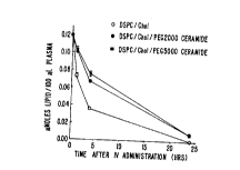
Figure 1A and-Figure iB illustrate graphically the
circulation lifetimes of the PEG modified ceramide liposomes
of the invention. Figure 1A shows plasma clearance of 100 nm
liposomes prepared of distearylphosphatidylcholine
(DSPC) /Cholesterol (Chol) (55:45 mol$; open circles),
DSPC/Chol/PEG200oCeramide (50:45:5 mol$; filled circles), and
DSPC/Chol/PEG5000Ceramide (50:45:5 mol$; filled squares).
Figure iB shows plasma clearance of 100 nm liposomes prepared
of DSPC/Chol (55:45 mol$; open circles), Sphingomyelin
(SM)/Chol (55:45 mol$; filled circles), and
Sphingomyelin/Chol/PEG2000Ceramide (50:45:5 mol$; open
squares).
Figure 2 graphically shows that the incorporation of
PEG modified ceramide into liposomal vincristine formulations
does not adversely affect drug retention characteristics.
Vincristine retention by Sphingomyelin/Chol (55:45 mol$;
filled circles) liposomes within the circulation is not
affected by incorporation of PEG200oCeramide (50:45:5 mol$;
open squares). Vincristine retention is also shown for
DSPC/Chol (55:45 mol$; open circles) and SM/Chol/PEG-PE
(Phosphatidylethanolamine) (50:45:5 mol$; filled squares).
Figure 3 shows the lipid circulation half-life
(T1/2). values (in hours) of various SM/cholesterol liposomes,
including those containing PEG2000-ceramides of various fatty
amide chain lengths and those containing PEG2000-DSPE
(distearolyphosphatidylethanolamine).
Figure 4 illustrates the circulation half-life
values (Tl/Z) (in hours) of the vincristine/lipid ratios
(vincristine retention) for liposomes, containing vincristine
with various PEG2000-ceramides, as well as PEG2000-DSPE.
Figure 5 presents the circulation half-life values
(T1/2) (in hours) of vincristine-containing liposomes.
2201120
WO 96/10391 PCT/CA95/00556
6
Figure 6 graphically shows the effect of increasing
concentrations of PEG-Ceramide (C20) on biodistribution of
liposomes in the blood and liver. 3H-labeled liposomes
composed of DOPE (dioleoylphosphatidylethanolamine), 15 mol%
DODAC (N,N-dioleoyl-N,N-dimethylammonium chloride) and the
indicated concentrations of PEG-Ceramide (C20) were injected
i.v. into mice. Biodistribution was examined at 1 hour after
injection, and the data were expressed as a percentage of the
injected dose in the blood (upper panel) and liver (lower
panel) with SD (standard deviation) (n=3).
Figure 7 graphically illustrates the effect of
increasing concentrations of DODAC on the biodistribution of
liposomes in the blood. 3H-labeled liposomes composed of
DOPE, 10 (open squares) or 30 (open triangles) mol% PEG-
Ceramide (C20), and the indicated concentration of DODAC were
injected i.v. into mice. Biodistribution was examined at
1 hour after injection, and the data were expressed as a
percentage of the injected dose in the blood.
Figure 8 graphically shows the liposome levels in
the blood and liver at different times after injection. 3H-
labeled liposomes composed of DOPE/DODAC (85:15 mol/mol) (open
circles with 0$ PEG-Ceramide (C20)), DOPE/DODAC/PEG-Ceramide
(C20) (75:15:10 mol/mol/mol) (open squares with 10% PEG-
Ceramide (C20)), and DOPE/DODAC/PEG-Ceramide (C20) (55:15:30
mol/mol/mol) (open triangles with 30% PEG-Ceramide (C20)) were
injected i.v. into mice. Biodistribution was examined at
indicated times, and the data were expressed as a percentage
of the injected dose in the blood (upper panel) and in the
liver (lower panel) with SD (n=3).
Figure 9 graphically illustrates the fusion of
PEG2000-DMPE and PEG2000-Ceramide (C14:0) containing vesicles
with an anionic target.
DETAILED DESCRIPTION OF THE PREFERRED EMBODIMENT
The PEG-modified ceramide lipids of Formula I
enhance the properties of liposomes by increasing the
circulation longevity or lifetime of the liposome; preventing
aggregation of the liposomes during covalent protein coupling,
2201120
WO 96/10391 PCT/CA95/00556
7
such as for targeting; preventing aggregation of liposomes
incorporating targeting moieties or drugs, such as antibodies,
and DNA; promoting drug retention within the liposome; and/or
increasing bilayer or other stability of the liposome when low
pH is required for encapsulation of the bioactive agents.
These PEG-Ceramide lipids also reduce leakage due to
hydrolysis of the fatty acyl chains of the liposome bilayer
and are more stable than other lip~i forms.
Definitions
As used herein, the term "alkyl" denotes branched or
unbranched hydrocarbon chains, such as, e.g., methyl, ethyl,
n-propyl, iso-propyl, n-butyl, sec-butyl, iso-butyl, tert-
butyl, and 2-methylpentyl. These groups may be optionally
substituted with one or more functional groups which are
attached commonly to such chains, such as, e.g., hydroxyl,
bromo, fluoro, chloro, iodo, mercapto or thio, cyano,
alkylthio, heterocycle, aryl, heteroaryl, carboxyl,
carbalkoyl, alkyl, alkenyl, nitro, amino, alkoxyl, amido, and
the like to form alkyl groups such as trifluoromethyl, 3-
hydroxyhexyl, 2-carboxypropyl, 2-fluoroethyl, carboxymethyl
and cyanobutyl and the like.
The term "alkylene" refers to divalent alkyl as
defined above, e.g., methylene (-CH2-), propylene
(-CH2CH2CH2-), chloroethylene (-CHCICH2-), 2-thiobutene
(-CH2CH(SH)CH2CH2-), 1-bromo-3-hydroxyl-4-methylpentene
(-CHBrCH2CH(OH)CH(CH3)CH2-) and the like.
The term "alkenyl" denotes branched or unbranched
hydrocarbon chains containing one or more carbon-carbon double
bonds.
The term "alkynyl" refers to branched or unbranched
hydrocarbon chains containing one or more carbon-carbon triple
bonds.
The term "aryl" denotes a chain of carbon atoms
which form at least one aromatic ring having preferably
between about 6-14 carbon atoms, such as, e.g., phenyl,
naphthyl, indenyl, and the like, and which may be substituted
with one or more functional groups which are attached commonly
2201120
WO 96/10391 PCT/CA95/00556
8
to such chains, such as, e.g., hydroxyl, bromo, fluoro,
chloro, iodo, mercapto or thio, cyano, cyanoamido, alkylthio,
heterocycle, aryl, heteroaryl, carboxyl, carbalkoyl, alkyl,
alkenyl, nitro, amino, alkoxyl, amido, and the like to form
aryl groups such as biphenyl, iodobiphenyl, methoxybiphenyl,
anthryl, bromophenyl, iodophenyl, chlorophenyl, hydroxyphenyl,
methoxyphenyl, formylphenyl, acetylphenyl,
trifluoromethylthiophenyl, trifluoromethoxyphenyl,
alkylthiophenyl, trialkylammoniumphenyl, amidophenyl,
thiazolylphenyl, oxazolylphenyl, imidazolylphenyl,
imidazolylmethylphenyl and the like.
The term "acyl" denotes groups -C(O)R, where R is
alkyl or aryl as defined above, such as formyl, acetyl,
propionyl, or butyryl.
The term "alkoxy" denotes -OR-, wherein R is alkyl.
The term "amido" denotes an amide linkage: -C(O)NH-.
The term "amino" denotes an amine linkage: -NR-
wherein R is hydrogen or alkyl.
The term "carboxyl" denotes -C(O)O-, and the term
"carbonyl" denotes -C(O)-.
The term "carbonate" indicates -OC(O)O-.
The term "carbamate" denotes -NHC(O)O-, and the term
"urea" denotes -NHC(O)NH-.
The term "phosphoro" denotes -OP(O)(OH)O-.
Structure and Preparation of Lipid Compounds
The compounds of the invention are synthesized using
standard techniques and reagents. It will be recognized that
the compounds of the invention will contain various amide,
amine, ether, thio, ester, carbonate, carbamate, urea and
phosphoro linkages. Those of skill in the art will recognize
that methods and reagents for forming these bonds are well
known and readily available. See, e.g., March, ADVANCED ORGANIC
CHEMISTRY (Wiley 1992), Larock, COMPREHENSIVE ORGANIC TRANSFORMATIONS
(VCH 1989) ; and Furniss, VOGEL 'S TEXTBOOK OF PRACTICAL ORGANIC
CHEMISTRY 5th ed. (Longman 1989). It will also be appreciated
that any functional groups present may require protection and
deprotection at different points in the synthesis of the
2201120
WO 96/10391 PCT/CA95/00556
9
compounds of the invention. Those of skill in the art will
recognize that such techniques are well known. See, e.g.,
Green and Wuts, PROTECTIVE GROUPS IN ORGANIC SYNTHESIS (Wiley
1991).
A general sequence of reactions for forming the
compound of the invention is illustrated below in Reaction
Scheme I. As shown therein, ceramide derivative 1 is reacted
with the PEG derivative PEG-Y1-alk-RG. R1-R4 and Y1 have
their meanings as defined above. RG is a group which reacts
with X2 to form the desired linkage Y2 between PEG and the
ceramide derivative (i.e., -Y1-alk-Y2-). Thus, it will be
appreciated that the identities of RG and X2 will be
complementary to each other and defined in such a way as to
provide the desired linkage. For example, where RG is a
nucleophilic center, such as -SH, -OH, or -NH21 X2 may be
oxygen derivatized to form a good leaving group, such as -OTs
where Ts represents the tosyl group, or halogen. Conversely,
X2 may be a nucleophilic center, e.g., -SH, -OH, or -NH2, and
RG a group which is reactive toward nucleophilic attack, e.g.,
carboxyl activated with dicyclohexylcarbodiimide (DCC) or acyl
chloride (-COCl). By suitable choice of RG and X2, the
desired amido, amine, ether, ester, thioether, carboxyl,
carbamate, carbonyl, carbonate, urea or phosphoro coupling
between the linker and the ceramide may be obtained. Finally
any protecting groups, e.g., R8, remaining on the intermediate
2 are converted to form the desired PEG-Ceramide derivative 3.
2201120
WO 96/10391 PCT/CA95/00556
O
R2
Ri N R4
X2
R8X1 -'R3
PEG-Y1-alk-RG
O
2
Rl R,, N R4
PEG -Y
R$X1 2
O
2
R1R ~N R4
PEG -Y
\
R5X1 R3 3
Reaction Scheme I
An exemplary synthesis of the PEG-Ceramide lipids of
the invention wherein Y1 and Y2 are carboxyl is illustrated
5 below in Reaction Scheme II. To eliminate the potential
problem of crosslinkage formation, PEG is capped at one end by
an unreactive group such as methoxy or ethoxy. The second
hydroxy group at the other terminal of the PEG molecule is
either activated with a suitable reagent such as cyanuric
10 acid, 1,1'-carbonyldiimidazole (CDI) or tresyl halide.
Alternatively the terminal hydroxyl group may first be
converted to a derivative that can be readily reacted with
ceramide in the presence of appropriate condensation reagents,
such as the succinate or amine. In other alternative methods,
the hydroxy groups on ceramide can be selectively activated
WO 96/10391 2201120 PCT/CA95/00556
11
for conjugation with PEG, or the two compounds can be linked
in a concerted coupling reaction by established coupling
procedures.
In the example shown, the primary hydroxyl group of
ceramide [available commercially from Sigma Chemical Company
(St. Louis, Missouri) and Avanti Polar Lipids Inc. (Alabaster,
Alabama)] is reacted with a hydroxyl protecting group of the
type which favors reaction at primary alcohols over secondary
and tertiary alcohols. Preferred protecting groups are those
which are considered sterically hindered, such trityl chloride
(TrCl) which comprises three phenyl rings attached to a
central carbon atom. However, other protecting groups are
known in the art (see, Green and Wuts supra). This reaction
is performed using standard techniques and conditions.
Following the protection of the C1 hydroxyl group,
the secondary alcohol at C3 is protected with a second
protecting group. The second protecting group should be one
which is reactive towards more hindered secondary alcohols,
but which is not removed under conditions effective to remove
the protecting group blocking the C1 alcohol. A preferred
protecting group is benzyl (Bn). Again, other suitable
protecting group combinations will be apparent to those of
skill in the art.
Once both of the hydroxyl groups are protected, the
C1-OH protecting group is removed under conditions which do
not affect the protecting group at the C3 alcohol. The free
hydroxyl function is then reacted with the PEG derivative
Me(PEG)OC(O)CH2CH2CO2H with dicyclohexylcarbodiimide (DCC) and
4-N,N'-dimethylaminopyridine (DMAP) to form the desired PEG-
Ceramide derivative.
The protecting group at C3 can be removed, if
desired, to permit other reactions at this site to obtain
other substituent groups.
2201120
WO 96/10391 PCT/CA95/00556
12
H NHC(O)R4
HO
H H
Trc1
H NHC (O) R 4
T=O
g H
BnCl
H NHC(O)R4
TrO
Bn "H
1. H+
2. Me(PEG)OC(0)CH2CH2COZH/DCC/DMAP
H NHC(O)R4
Me (PEG) OC (0) CH 2CH2C (0) O
DBn
Reaction Scheme II
In another approach, shown in Reaction Scheme III
below, Y1 is a carboxyl ester group -OC(O)- and Y2 is an amido
-C(O)NH-. As shown in the scheme, the 1-amino analog of
ceramide can be prepared by derivitization of the C1 hydroxyl
group first to the corresponding C1 alkyl sulfonate (e.g.,
methyl sulfonate or 2,2,2-trifluoroethanesulfonate). The
latter is converted to the amino analog directly with ammonia
or through an azide intermediate as shown. The 1-amino-
ceramide is then coupled to the N-hydroxysuccinamide (NHS)
ester of MePEG-S to form a MePEG-S-ceramide with an amide
linkage.
WO 96/10391 2201120 PCT/CA95/00556
13
H NHC (O) R 4
MeS020
H-H
NaN3
H NHC(O)R4
N3
N"3
OH 11
H NHC(O)R4
H2N
OH
Me (PEG) OC (O) C 2H4CO2H
DCC/NHS
-OC(0)C2H4C(O)O(PEG)Me
0
H H NHC (0) R 4
Me (PEG) OC (0) C 2H4C (0)-- N
OH ~H
Reaction Scheme III
WO 96/10391 2201120 PCT/CA95/00556
14
Alternatively, the group Y may be -NR7-, where R7 is
hydrogen, alkyl, acyl, or aryl; or Y may be -0- or -S-. Such
embodiments may be formed using well known methods and
reagents. For example, the embodiment wherein Y is -NH- can
be made by the synthesis pathway shown in Reaction Scheme IV
below. There, the 1-mesyl-ceramide described above is reacted
with the amino analog of (MePEG-NH2) to form the desired
MePEG-Ceramide conjugate having an amino linkage.
H NHC(O)R4
MeSO20
H
H
Me PEG-OH
MSC1/EtjN
MePEG-OSO2Me
NH4OH
MePEG-NHz
H NHC (O) R 4
MePEGNH
Reaction Scheme IV
Both the C1 and C3 hydroxy functions in ceramide can be
activated with a reagent such as CDI to form the corresponding
bis-imidazolyl formate. The latter is then reacted with the
amino group of MePEG-NH2 to form a conjugate with two MePEG
WO 96/10391 2201120 PCT/CA95/00556
molecules bonded to one ceramide. Either one or two PEG
molecules can be selected to attach to each ceramide, allowing
a more flexible arrangement for introducing specific
properties to a liposomal system.
5 The group Y = Y1-alk-Y2 can be formed from readily
available starting materials using known techniques.
Preferred embodiments include those wherein Y1 and Y2 are both
carbonyl (-C(O)-) or where one of Y1 or YZ is carbonyl and the
other is amido (-C(O)NH-). These groups can be formed from
10 commercially available diacids, such as malonic acid
(CH2(CO2H)2)1 succinic acid (H02CCHZCH2CO2H), glutaric acid
(HO2CCH2CH2CH2CO2H) and adipic acid (H02CCHZCH2CH2CH2CO2H) and
the like; as well as substituted diacids, such as tartaric
acid (HO2CCH(OH)CH(OH)CO2H), 3-methylglutaric acid
15 (HO2CCH2CH(CH3)CH2CO2H) and the like, using methods well known
in the chemical arts. Acyl derivatives, such as acyl
chlorides, e.g., 3-carbomethoxypropiponyl chloride
(C1C(O)C2H4CO2CH3), and amides corresponding to these
compounds are available commercially or can be formed using
known procedures.
PEG is a linear, water-soluble polymer of ethylene
oxide repeating units with two terminal hydroxyl groups. PEGs
are classified by their molecular weights; for example,
PEG 2000 has an average molecular weight of about 2,000
daltons, and PEG 5000 has an average molecular weight of about
5,000 daltons. PEGs are commercially available from Sigma
Chemical Co. and other companies and include:
monomethoxypolyethylene glycol (MePEG-OH),
monomethoxypolyethylene glycol-succinate (MePEG-S),
monomethoxypolyethylene glycol-succinimidyl succinate (MePEG-
S-NHS), monomethoxypolyethylene glycol-amine (MePEG-NH2),
monomethoxypolyethylene glycol-tresylate (MePEG-TRES), and
monomethoxypolyethylene glycol-imidazolyl-carbonyl (MePEG-IM).
The attachment of PEG to the linker Y may be
performed using methods and materials known in the art.
Generally, a hydroxyl or amino moiety of the PEG group, is
reacted with suitable derivative of Y so as to form the
desired coupling. For example, reaction of a free hydroxyl
2201120
WO 96/10391 PCT/CA95/00556
16
functionality of MePEG-OH with an acyl chloride derivative,
such as 3-carbomethoxypropiponyl chloride (C1C(O)C2H4CO2CH3)1
available commercially from Aldrich Chemical Co., Milwaukee,
Wisconsin, provides Me(PEG)-OC(O)C2H4CO2CH3. The methyl ester
can be further derivatized, e.g., to the acyl chloride or
amide, using standard procedures. Alternatively,
Me(PEG)OC(O)CH2CH2CO2H may be formed from Me(PEG)-OH and
succinic anhydride as shown below:
0
MePEG- OH 010 MePEG- OC (0) CH2CH2C02H
0
Still other methods will be apparent to those of skill in the
art.
To couple PEG directly to the ceramide, the hydroxy
function in MePEG can be directly activated with reagent such
as CDI to form the corresponding imidazolyl formate. The
latter is then reacted with a nucleophile, such as one or both
alcohol functions of ceramide, to form a conjugate with a
carbonate linkage. Alternatively, coupling of the MePEG
imidazolyl formate with 1-aminoceramide will result in the
formation of a MePEG-Ceramide adduct with a carbamate linkage.
Commercial ceramides, which are N-acyl fatty acids
of sphingosines, may be obtained by phospholipase C cleavage
of the phosphocholine in the respective sphingomyelin
precursors, which are extracted from egg yolks and brain
tissue. The sphingomyelin lipids differ in the composition of
the fatty amide chains, such as in the carbon chain length and
the number of double bonds. The following ceramides are
commercially available from Sigma Chemical Co. and Avanti
Polar Lipids Inc.: (1) Type III: from bovine brain
(approximately 99%); (2) Type IV: bovin brain (approximately
99%); (3) from brain (approximately 99%); and (4) from egg
(approximately 99%). The fatty amide chains differ in
composition based upon the source of the sphingomyelin, as
WO 96/10391 2201120 PCT/CA95/00556
17
shown in Table I:
Table I
FATTY ACID CONTENT OF TISSUE DERIVED SPHINGOMYELIN
Fatty Acid Egg Sphingomyelin Brain Sphingomyelin
16:0 77.70 2.38
18:0 7.44 57.99
20:0 1.83 6.08
22:0 3.98 9.16
24:0 1.86 7.04
24:1 2.80 14.71
A wide variety of ceramide derivatives may be
synthesized from common starting materials using known
techniques. For example, starting from commercially available
erythritol (Aldrich, Milwaukee, Wisconsin), any ceramide
derivative may be synthesized as illustrated in Reaction
Scheme V below. Selective protect?on of erythritol using
known methods provides the startir material shown in Reaction
Scheme V, wherein the C1 and C3 carbons are protected as the
benzyl (Bn) derivatives, the C2 carbon is protected as the
methylmethoxy (MOM) ether and the C4 carbon is protected as
the 3,4-dimethoxybenzyl (DMPM) ether derivative. Selective
removal of the DMPM group using dichlorodicyanoquinone (DDQ)
provides the corresponding alcohol which can be oxidized using
standard methods to form the aldehyde as shown (see, e.g.,
Larock, supra).
Reaction of the aldehyde with the Wittig reagent
Et3P+C14H29Br- provides the trans olefin preferentially.
Alternatively, reaction with the triphenylphosphine derivative
Ph3P+C14H29Br- provides the cis olefin predominantly. Again,
other methods of olefin formation will be apparent to those of
skill in the art. Removal of the MOM protecting group,
followed by conversion of the alcohol using sodium azide
(NaN3) and lithium aluminum hydride (LiAlH4) provides the
desired amine which is reacted with an acyl chloride to
produce the amide shown. Reaction of the amide with boron
WO 96/10391 2201120 PCT/CA95/00556
18
trichloride (BC13) in methylene chloride (CH2C12) using a
temperature gradient from -78 C to 0 C, followed by reaction
with methanol (MeOH) at -78 C, provides the desired diol.
Other equivalent methods of synthesis will be apparent to
those of skill in the art. Additional information regarding
the synthesis of sphingolipids and optically active ceramides
can be found in Schmidt, et al. ANGEW. CHEM. INT. Ed. Engl
(1987) 26:793; Kiso, et al. J. CARBOHYDR. CHEM. (1986) 5:335; and
Nicolaou, et al. J. AMER. CHEM. Soc. (1988) 110:7910.
Ceramides of varying fatty amide chain lengths also
can be prepared by reacting the amine of Scheme V with various
acyl chlorides [R4C(O)C1] or other acyl or acid derivatives,
whereby the carbon chain length is based upon the particular
acyl group used. Typically and preferably, the carbon chain
length is from about 8 to about 24, without any double bonds
present, e.g., an alkyl chain. Most preferred are those
ceramides designated as 20:0, which designates a 20 carbon
length chain with no double bonds, i.e., a completely
saturated C20 alkyl as the fatty amide chain. Alternatively,
ceramides with specific acyl chains of homogenous composition
can be prepared by conjugation of a suitably activated
carboxylic compounds, such as N-hydroxysuccinimide (NHS) ester
of fatty acid, with the amino function of D-sphingosine. For
example, an acyl chloride of eicosanoic acid (also known as
arachidic acid) will provide a chain 20 carbons (C20) in
length for the resulting amide side chain. Other acids
preferably include: octanoic acid (also known as caprylic
acid) for C8; myristic acid for C14; palmitic acid (also known
as hexadecanoic acid) for C16; and tetracosanoic acid (also
known as lignoceric acid) for C24. Ceramides with fatty amide
chain lengths of 14 to 20 carbons are especially preferred.
Most preferred are those 14 or 20 carbons in length. (It is
understood that R4 is one carbon shorter in length than the
starting acyl chloride or acid.)
2201120
WO 96/10391 PCT/CA95/00556
19
OMOM OMOM
BnO DDQ BnO
ODMPM OH
OBn OBn
(O]
MOM 0
BnO
H
Bn
Et3P+C14Hz9Br
MOM
BnO OBn
1. Me3SiBr/CHZC12/0 C
2. MaC1/Py/0 C
3. vaN3/DMF/0
4. LiAlH4
NH2
Bn0
OBn
R4C(O)C1
NHC(0)R4
Bn0
OBn 1. BC13/CH2Cl2/-78 to 0 C
2. MeOH/-78 C
NHC(0)R4
HO
OH
Reaction Scheme V
2201120
WO 96/10391 PCT/CA95/00556
Liposome Preparation
After the lipids of Formula I are prepared, they can
be utilized in liposome structures incorporating or entrapping
one or more bioactive agents, wherein one or more of the lipid
5 compounds comprise the liposome. For example, the fatty amide
chain can have various lengths on the ceramide portion of the
lipid, and a mixture of the various resulting lipid compounds
forms the desired liposome. In addition, non-PEG ceramide
lipids can be used to construct the liposome in conjunction
10 with the lipids of Formula I.
A variety of methods are available for preparing
liposomes as described in, e.g., Szoka et al., 9 ANN. REV.
BIOPHYs. BIOENG. 467 (1980) ; U.S. Pat. Nos. 4,235,871,
4,501,728, 4,837,028; the text LiPosorsEs Ch. 1 (supra) and Hope
15 et al., 40 CHEM. PHxs. LIP. 89 (1986). One method produces
multilamellar vesicles of heterogeneous sizes. In this
method, the vesicle-forming lipids are dissolved in a suitable
organic solvent or solvent system and dried under vacuum or an
inert gas to form a thin lipid film. Alternatively, the
20 lipids may be dissolved in an organic solvent such as tert-
butyl alcohol or benzene:methanol (95:5 v/v) and lyophilized
to form a homogeneous lipid mixture, which is in a more easily
hydrated powder-like form. The dry lipid mixture is covered
with an aqueous buffered solution and allowed to hydrate,
typically over a 15-60 minute period with agitation. The size
distribution of the resulting multilamellar vesicles can be
shifted toward smaller sizes by hydrating the lipids under
more vigorous agitation conditions. Full hydration of the
lipids may be enhanced by freezing in liquid nitrogen and
thawing to about 50 C. This cycle is usually repeated about
five times.
Following liposome preparation, the liposomes may be
sized to achieve a desired size range and relatively narrow
distribution of liposome sizes. A size range of about 0.05-
0.20 microns allows the liposome suspension to be sterilized
by filtration through a conventional filter, using typically a
0.22 micron filter. The filter sterilization method can be
carried out on a high through-put basis if the liposomes have
WO 96/10391 2201 12O PCT/CA95/00556
21
been sized down to about 0.05-0.20 microns.
Several techniques are available for sizing
liposomes, such as the sizing method described in U.S. Pat.
No. 4,737,323. For example, sonicating a liposome suspension
either by bath or probe sonication produces a progressive size
reduction down to small unilamellar vesicles less than about
0.05 microns in size. Homogenization is another method which
relies on shearing energy to fragment large liposomes into
smaller ones. In a typical homogenization procedure,
multilamellar vesicles are recirculated through a standard
emulsion homogenizer until selected liposome sizes, typically
between about 0.01 and 0.5 microns, are observed. In both
methods, the particle size distribution can be monitored by
conventional laser-beam particle size discrimination.
Extrusion of liposome through a small-pore
polycarbonate membrane or an asymmetric ceramic membrane is
also an effective method for reducing liposomes to a
relatively well-defined size distribution. Typically, the
suspension is cycled through the membrane one or more times
until the desired liposome size distribution is achieved. The
liposomes may be extruded through successively smaller-pore
membranes, to achieve a gradual reduction in liposome size.
For use in the present invention, liposomes having a size of
from about 0.05 microns to about 0.20 microns are preferred.
Liposome preparations are also described by Deamer et al., in
Liposomes (supra) LIPOSOME PREPARATIONS: METHODS AND MECHANISMS.
Liposome size distributions also may be determined
by quasi-elastic light scattering techniques. See Bloomfield,
10 Ann. Rev. Biophys. Bioeng. 421 (1981).
Use of Liposomes as Delivery Vehicles
The liposomes prepared by using the lipid compounds
of this invention can be labeled with markers that will
facilitate diagnostic imaging of various disease states
including tumors, inflamed joints or lesions. Typically,
these labels will be radioactive markers, although fluorescent
labels can also be used. The use of gamma-emitting
radioisotopes is particularly advantageous as they easily can
2201120
WO 96/10391 PCT/CA95/00556
22
be counted in a scintillation well counter, do not require
tissue homogenization prior to counting, and can be imaged
with gamma cameras.
Gamma- or positron- emitting radioisotupes are
typically used, such as 99Tc, 24Na, 51Cr, 59Fe, 67Ga, 86Rb,
111In, 125I, and 195Pt as gamma-emitting; and such as 68Ga,
82Rb, 22Na, 75Br, 1221 and 18F as positron-emitting.
The liposomes also can be labelled with a
paramagnetic isotope for purposes of in vivo diagnosis, as
through the use of magnetic resonance imaging (MRI) or
electron spin resonance (ESR). See, for example, U.S. Pat.
No. 4,728,575.
Liposomes are a valuable system for the controlled
delivery of drugs. As discussed earlier, liposomes formulated
from PEG-lipids are especially advantageous, since they are
more stable and have an increased half-life in circulation
over conventional liposomes. Using liposomes as drug carriers
allows more control of the site or rate of release of the
drug, enabling more precision to be obtained in regulating the
blood and organ levels of drug and/or its metabolites. Thus,
drug dosages needed to produce clinical effects can be reduced
which in turn reduces toxicity. Toxicity concerns are
particularly valid in cancer chemotherapy where the dose
levels required for beneficial effects and the doses that
result in significant toxicity are very close. Thus, for
cancer chemotherapy the use of liposome carriers for antitumor
drugs can provide significant therapeutic advantages.
Depending on the capture volume within the liposome
and the chemical and physical properties of the bioactive
agents, compatible bioactive agents can be simultaneously
encapsulated in a single liposome. Simultaneous delivery of
two or more synergistic drugs in this manner will ensure the
delivery of these drugs to the same location in the body and
maintain the drugs in close proximity to act together, thus
greatly facilitating therapy.
A wide variety of bioactive agents, pharmaceutical
substances, or drugs can be encapsulated within the interior
of the relatively impermeable bilayer membranes of the
2201120
WO 96/10391 PCT/CA95/00556
23
liposomes where these substances can be protected from the
environment during transit to their target areas. These
substances include antitumor agents, antibiotics,
immunomodulators, anti-inflammatory drugs and drugs acting on
the central nervous system (CNS). Especially preferred
antitumor agents include actinomycin D, vincristine,
vinblastine, cystine arabinoside, anthracyclines, alkylative
agents, platinum compounds, antimetabolites, and nucleoside
analogs, such as methotrexate and purine and pyrimidine
analogs. Considering the preferred uptake of intravenously
injected liposomes by the bone marrow, lymphoid organs, liver,
spleen and lungs, and the macrophage cell, neoplasms and other
diseases involving these organs can be effectively treated by
PEG-derived liposome-entrapped drug. (See Daoud et al.,
"Liposomes In Cancer Therapy", 3 ADv DRUG DELIVERY REVIEWS 405-
418, 1989.)
Another clinical application of liposomes is as an
adjuvant for immunization of both animals and humans. Protein
antigens such as diphtheria toxoid, cholera toxin, parasitic
antigens, viral antigens, immunoglobulins, enzymes,
histocompatibility antigens can be incorporated into or
attached onto the liposomes for immunization purposes.
Liposomes are also particularly useful as carriers
for vaccines that will targeted to the appropriate lymphoid
organs to stimulate an immune response.
Liposomes have been used as a vector to deliver
immunosuppressive or immunostimulatory agents selectively to
macrophages. In particular, glucocorticoids useful to
suppress macrophage activity and lymphokines that activate
macrophages have been delivered in liposomes.
Liposomes with targeting molecules can be used to
stimulate or suppress a cell. For example, liposomes
incorporating a particular antigen can be employed to
stimulate the B cell population displaying surface antibody
that specifically binds that antigen. Similarly, PEG-
stabilized liposomes incorporating growth factors or
lymphokines on the liposome surface can be directed to
stimulate cells expressing the appropriate receptors for these
2201120
WO 96/10391 PCT/CA95/00556
24
factors. Such an approach can be used for example, in
stimulating bone marrow cells to proliferate as part of the
treatment of cancer patients following radiation or
chemotherapy which destroys stem cells and actively dividing
cells.
Liposome-encapsulated antibodies can be used to
treat drug overdoses. The tendency of liposomes having
encapsulated antibodies to be delivered to the liver has a
therapeutic advantage in clearing substances such as toxic
agents from the blood circulation. It has been demonstrated
that whereas unencapsulated antibodies to digoxin caused
intravascular retention of the drug, encapsulated antibodies
caused increased splenic and hepatic uptake and an increased
excretion rate of digoxin.
Liposomes comprising PEG-lipids also find utility as
carriers in introducing lipid or protein antigens into the
plasma membrane of cells that lack the antigens. For example,
histocompatibility antigens or viral antigens can be
introduced into the surface of viral infected or tumor cells
to promote recognition and killing of these cells by the
immune system.
In certain embodiments, it is desirable to target
the liposomes of the invention using targeting moieties that
are specific to a cell type or tissue. Targeting of liposomes
using a variety of targeting moieties, such as ligands, cell-
surface receptors, glycoproteins, and monoclonal antibodies,
has been previously described. See U.S. Pat. Nos. 4,957,773
and 4,603,044. The targeting moieties can comprise the entire
protein or fragments thereof.
Targeting mechanisms generally require that the
targeting agents be positioned on the surface of the liposome
in such a manner that the target moiety is available for
interaction with the target; for example, a cell surface
receptor. The liposome is designed to incorporate a connector
portion into the membrane at the time of liposome formation.
The connector portion must have a lipophilic portion that is
firmly embedded and anchored into the membrane. It must also
have a hydrophilic portion that is chemically available on the
CA 02201120 2006-03-28
aqueous surface of the liposome. The hydrophilic portion is
selected so as to be chemically suitable with the targeting
agent, such that the portion and agent form a stable chemical
bond. Therefore, the connector portion usually extends out
5 from the liposome's surface and is configured to correctly
position the targeting agent. In some cases it is possible to
attach the target agent directly to the connector portion, but
in many instances, it is more suitable to use a third molecule
to act as a "molecular bridge." The bridge links the
10 connector portion and the target agent off of the surface of
the liposome, making the target agent freely available for
interaction with the cellular target.
Standard methods for coupling the target agents can
be used. For example, phosphatidylethanolamine, which can be
15 activated for attachment of target agents, or of derivatized
lipophilic compounds, such as lipid-derivatized bleomycin, can
be used. Antibody-targeted liposomes can be constructed
using, for instance, liposomes that incorporate protein A.
See Renneisen et al., 265 J. Biol. Chem. 16337-16342 (1990)
20 and Leonetti et al., 87 Proc. Nat1. Acad. Sci. (USA) 2448-2451
(1990). Other examples of antibody conjugation are disclosed
in WO 96/10585.
Examples of targeting moieties also can include
25 other proteins, specific to cellular components, including
antigens associated with neoplasms or tumors. Proteins used
as targeting moieties can be attached to the liposomes via
covalent bonds. See Heath, Covalent Attachment of Proteins to
Liposomes, 149 Methods in Enzymology 111-119 (Academic Press,
Inc. 1987). Other targeting methods include the biotin-avidin
system.
In some cases, the diagnostic targeting of the
liposome can subsequently be used to treat the targeted cell
or tissue. For example, when a toxin is coupled to a targeted
liposome, the toxin can then be effective in destroying the
targeted cell, such as a neoplasmic cell.
Once the encapsulated bioactive agents or the
liposomes themselves are taken up by the cell, the bioactive
WO 96/10391 2201120 PCT/CA95/00556
26
agents also can be targeted to a specific intracellular site
of action if target recognizing moieties are incorporated into
the agent. For example, protein agents to be delivered to the
nucleus may comprise a nuclear localization signal sequence
recombinantly engineered into the protein or the signal
sequence may be on a separate protein or peptide covalently
attached to the primary protein. Likewise, non-protein drugs
destined for the nucleus may have such a signal moiety
covalently attached. Other target recognizing moieties that
can be recombinantly engineered into or covalently attached to
protein components to be delivered by liposomes include
ligands, receptors and antibodies or fragments thereof.
The present invention also provides a kit for
preparing labeled liposomes. The kit will typically be
comprised of a container that is compartmentalized for holding
the various elements of the kit. One compartment can contain
the materials for preparing the label just prior to use. A
second compartment can contain the liposomes with or without a
pH buffer to adjust the composition pH to physiological range
of about 7 to about S. The liposomes also can be provided in
freeze-dried form for reconstitution at the time of use. Also
included within the kit will be other reagents and
instructions for use.
Liposomes comprising the lipid compounds of this
invention can be formulated as pharmaceutical compositions or
formulations according to standard techniques using acceptable
adjuvants or carriers. Preferably, the physiologically
pharmaceutical compositions are administered parenterally,
i.e., intravenously, subcutaneously, or intramuscularly.
Suitable formulations for use in the present invention are
found in REMINGTON 'S PHARMACEUTICAL SCIENCES (Mack Publishing Co.,
18th ed. 1990).
Preferably, the compositions are administered
intravenously. Therefore, this invention provides for
compositions for intravenous administration which comprise a
solution of liposomes suspended in a physiologically-
acceptable adjuvant or carrier, preferably an aqueous carrier,
such as water, buffered water, isotonic saline, and the like.
2201120
WO 96/10391 PCT/CA95/00556
27
The compositions may be sterilized by conventional, well-known
sterilization techniques, or may be sterile filtered. The
resulting aqueous solutions may be packaged for use as is, or
lyophilized; the lyophilized preparation being combined with a
sterile aqueous solution prior to administration. The
compositions can contain pharmaceutically-acceptable auxiliary
substances as required to approximate the appropriate
physiological conditions, such as pH adjusting and buffering
agents, tonicity adjusting agents, wetting agents, and the
like. Such agents include sodium acetate, sodium lactate,
sodium chloride, potassium chloride, calcium chloride,
sorbitan monolaurate, triethanolamine oleate, and the like.
The concentration of liposomes useful in
pharmaceutical compositions can range from about 0.05%,
usually about 2-5%, or as much as about 10-30% by weight of
the composition and the range of concentration is selected in
accordance with the mode of administration and bioactive agent
contained within the liposomes.
Since the present liposomes made from PEG-Ceramide
lipids are less susceptible to hydrolysis, they have a
prolonged half-life resulting in prolonged circulation.
Additionally, the liposome pharmaceutical composition can
include lipid-protective agents that protect the liposomes
against free-radical and lipid-peroxidative damage upon
storage. Such protective agents include alpha-tocopherol and
water-soluble, iron-specific chelators, such as ferrioxamine.
Use of Lipids or Lipid-Based Carriers as Delivery Vehicles
Cationic lipids may be used in the delivery of
therapeutic genes or oligonucleotides intended to induce or to
block production of some protein within the cell. Nucleic
acid is negatively charged and must be combined with a
positively charged entity to form a lipid complex suitable for
formulation and cellular delivery.
Cationic lipids have been used in the transfection
of cells in vitro and in vivo (Wang C-Y, Huang L. pH-sensitive
immunoliposomes mediate target cell-specific delivery and
controlled expression of a foreign gene in mouse. Pxoc. NATL.
WO 96/10391 2201120 PCT/CA95/00556
28
ACAD. Sci USA, 1987; 84:7851-7855 and Hyde SC, Gill DR. Higgins
CF, et al. Correction of the ion transport defect in cystic
fibrosis transgenic mice by gene therapy. NATURE. 1993;362:250-
255.) The efficiency of this transfection has often been less
than desired, for various reasons. One is the tendency for
cationic lipids complexed to nucleic acid to form
unsatisfactory carriers. These carriers are improved by the
addition of the PEG-modified ceramide lipids of the present
invention. The addition of PEG-modified ceramide lipids
prevents particle aggregation and provides a means for
increasing circulation lifetime and increasing the delivery of
the lipid-nucleic acid particles to the target cells.
Moreover, it has been found that cationic lipid carrier
systems fuse more readily with the target cells and, thus, the
addition of negatively charged PEG-PE lipids (e.g., PEG2000-
DMPE), while preventing the aggregation and self-fusion of the
cationic carrier plasmid systems, can introduce additional
electrostatic interaction between the anionic PEG-PE and
cationic lipid. Such interaction effectively retains the PEG-
PE on the carrier system and, thus, maintains the steric
stabilization effect and inhibits the fusion of the cationic
carrier system with the target cells. The neutral PEG-
modified ceramide lipids do not have such electrostatic
interaction with the cationic carrier components and, thus,
they can exchange out of the system according to the strength
of the affinity between the specific ceramide lipid and the
other hydrophobic components in the system. This is a
distinct advantage the PEG-modified ceramide lipids have over
PEG-PE in the formation of programmable fusogenic cationic
carrier systems (See, Example 12, infra).
Cationic lipids useful in producing lipid-based
carriers for gene and oligonucleotide delivery are LIPOFECTIN
(US patents US 4,897,355; 4,946,787; and 5,208,036 by Eppstein
et al.) and LIPOFECTACE (US patent 5,279,883 by Rose). Both
agents, as well as other transfecting cationic lipids, are
available from Life Technologies, Inc. in Gaithersburg,
Maryland.
The invention will be better understood by reference
2201120
WO 96/10391 PCT/CA95/00556
29
to the following examples, which are intended to illustrate
aspects of the invention, but the invention is not to be
considered as limited thereto.
Example 1
N-Eicosanovl-D-Sphinaosine jC20:0-Ceramidel
N-Hydroxysuccinimide (NHS) ester of eicosanoic acid
was synthesized using the procedure of Lapidot et al. (J.
Lipid Res., 1967, 8:142). 428 mg of the ester was added to a
solution of D-sphingosine (Avanti Polar Lipids, Inc.) (300 mg)
in anhydrous methylene chloride (CH2C121 16 ml) and
triethylamine (118 mg) with stirring under nitrogen (N2) at
C for 4 hours. Analysis by thin layer chromatography
(t.l.c.) (silica gel, CHC13:CH3OH:H20 - 65:25:4 v/v or
15 CHC13:CH3OH - 90:10 v/v) indicated most of the D-sphingosine
had reacted. [If necessary, another small portion of NHS-
ester of eicosanoic acid (20-30 mg) may be added to complete
the acylation of D-sphingosine]. The reaction mixture was
cooled in ice and diluted with CH2C12 (60 ml), H20 (30 ml) and
20 neutralized with 1N HC1. The CH2C12 layer was washed with H20
(2 X 30 ml) and dried (MgSO4) before evaporation to dryness in
vacuo. The residue was recrystallized twice from acetone to
give the pure product, N-eicosanoyl-D-sphingosine (428 mg), as
a white solid. T.l.c. showed a single spot and 1H-NMR
spectrum was consistent with the expected structure.
Esample 2
Monomethoxypolyethylene Glyco1200~-Succinate (MePEG2000=Si
Monomethoxypolyethylene glycol with an average
molecular weight of 2000 daltons (MePEG2000) (Sigma Chemical
Co.), (4g) dissolved in CH2C12 (30 ml) was treated with
succinic anhydride (600 mg), triethylamine (400 mg) and 4-
dimethylaminopyridine (DMAP) (250 mg), and stirred under N2 at
20 C for 16 hours. The reaction solution was diluted with
CH2C12 (60 ml), cooled in ice and H20 (50 ml) added. The
mixture was acidified with 1N HC1 and the organic layer
separated. The aqueous layer was further extracted with
CH2C12 (2 X 30 ml). The combined organic extracts were dried
WO 96/10391 2201120 PCT/CA95/00556
(MgSO4) and then evaporated to dryness. The crude product was
purified on a silica gel (G60) column eluted with a solvent
system of CH2C12 containing 2 to 8% methanol. Fractions
collected were analyzed by t.l.c. (silica gel, CiiC13:CH3OH-
5 88:12 v/v) and those containing the pure product (MePEG2000-S)
with Rf value of 0.4 were pooled and concentrated.
Trituration of the product with diethyl ether gave MePEG2000-S
as a white solid (3.2g).
10 Example 3
Monomethoxypolyethylene Glyco15000-Succinate IMePEG5000=-sl
The titled compound was prepared from
monomethoxypolyethylene glycol with an average molecular
weight of 5000 daltons (MePEG5000) (Sigma Chemical Co.) in a
15 similar procedure as described above for MePEG2000-S=
Example 4
1-0- ePEG2000-S)-(C20:0-Ceramide)
C20:0-Ceramide (60 mg), dicyclohexylcarbodiimide
20 (DCC) (28 mg) and DMAP (13 mg) were dissolved in warm
anhydrous CH2C12 (6 ml). MePEG2000-S (230 mg) in anhydrous
CH2C1Z (1 ml) was added dropwise to the above solution with
stirring under N2 at 25 C for 6 hours. The precipitated
dicyclohexylurea (DCU) was filtered off and the filtrate
25 concentrated in vacuo. Trituration of the solid residue with
diethyl ether removed most of the DCC, DMAP and unreacted
C20:0-ceramide. The resulting crude product was
chromatographed on a short silica gel column (G60) eluted with
CHZCI2:CH3OH-98:2 (v/v). Fractions containing the product
30 were combined and evaporated to dryness in vacuo. The
resulting solid was dissolved in distilled H20 (2 ml) and
dialysed at 4 C against distilled water overnight. The pure
product was obtained as a white powder (160 mg) by
lyophilization. T.l.c. (silica gel, CHCI3:CH3OH-90:10 v/v
showed a single spot (Rf 0.5). 1H-NMR spectrum of the product
was consistent with the structure of 1-0-(MePEG2000-S)-(C20:0-
ceramide). [PEG2000 Ceramide]
WO 96/10391 2201120 PCT/CA95/00556
31
Example 5
1-0- (MePEG2000-S ) - ( Egct Cerair ~ le ) [ PEGZOO0 Ceramide l
Egg ceramide (Avanti Po.Lar Lipids, Inc.) (108 mg),
DCC (48 mg), and DMAP (25 mg) were dissolved in warm anhydrous
CH2C12 (8 ml) . MePEG2000-S (460 mg) in anhydrous CH2C12 (2 ml)
was added dropwise to the above solution with stirring under
N2 at 25 C for 6 hours. The precipitated DCU was filtered off
and the filtrate concentrated in vacuo. Trituration of the
solid residue with diethyl ether removed most of the residual
reagents and small amount of unreacted egg ceramide. The
crude product was chromatographed on a short silica gel column
(G60) eluted with CH2C12:CH3OH-98:2 (v/v). Fractions
containing the product were combined and evaporated to dryness
in vacuo. The resulting solid was dissolved in distilled
water (2 ml) and dialysed at 4 C against distilled water
overnight. Lyophilization of the solution gave the pure
product as a white powder (338 mg). T.l.c. (silica gel,
CHC130H-90:10 v/v) showed a single spot (Rf 0.5) 1H-NMR
spectrum of the product was in agreement with the structure of
1-0-(MePEG2000-S)-(egg ceramide).
Example 6
1-0-(MePEG500o-S)-(egg Ceramide) [PEG5o0o Ceramidel
The titled compound was prepared from MePEG5000-S
(550 mg) and egg ceramide (54 mg) in a procedure similar to
that described above for 1-0-(MePEG2000-S) -(egg ceramide)
using DCC (28 mg) and DMAP (13 mg) as the condensation
reagents in anhydrous CH2C12 (6 ml). Similar purification by
column chromatography (silica gel) and dialysis gave the pure
product 1-0-(MePEG5000-S) -(egg ceramide) as a white powder
(310 mg).
Example 7
Plasma Clearance of Specific Liposomes
In this example, the plasma clearance for l00nm
liposomes prepared of Distearylphosphatidylcholine
(DSPC)/Cholesterol (Chol) (55:45 molt; open circles);
DSPC/Chol/PEG200oCeramide (50:45:5 molt; filled circles), and
2201120
WO 96/10391 PCT/CA95/00556
32
DSPC/Chol/PEG500oCeramide (50:45:5 molt; filled squares) was
determined. The results are shown in Figure lA. Lipid
mixtures were prepared in chloroform (CHC13) and subsequently
dried under a stream of nitrogen gas. The resulting lipid
film was placed under high vacuum for at least 2 hours prior
to hydration with 150 mM sodium chloride and 20 mM Hepes (pH
7.4) (Hepes buffered saline solution). Liposomes were then
prepared by extrusion through l00nm pore size filters using an
Extruder pre-heated to 65 C prior to extrusion. The resulting
liposomes exhibited a mean diameter of approximately 120nm.
These liposomes were diluted such that mice (female CD1) could
be given an i.v. dose of lipid equivalent to 50 moles/kg in
an injection volume of 200 l. At various time points
indicated in Figure 1A, blood samples were taken by nicking
the tail vein and collecting 25 l of blood into a EDTA
(ethylenediaminetetraacetic acid) coated capillary tube. The
amount of lipid in the resulting sample was determined by
measuring the amount of 3(H]-cholesteryl hexadecyl ether
present. This non-exchangeable, non-metabolizable lipid
marker was incorporated into the liposomes prior to formation
of the lipid film.
Example 8
Plasma Clearance of Specific Libosomes
This example illustrates the plasma clearance for
l00nm liposomes prepared of DSPC/Chol (55:45 mol$; open
circles), Sphingomyelin/Chol (55:45 mol$; filled circles), and
Sphingomyelin/Chol/PEG2000Ceramide (50:45:5 molt; open
squares). The results are presented in Figure 1B. Lipid
mixtures were prepared in chloroform (CHC13) and subsequently
dried under a stream of nitrogen gas. The resulting lipid
film was placed under high vacuum for at least 2 hours prior
to hydration with a 300 mM citrate buffer (pH 4.0). Liposomes
were then prepared by extrusion through 100nm pore size
filters using an Extruder pre-heated to 65 C prior to
extrusion. The resulting vesicles were diluted with 150 mM
NaCl, 20 mM Hepes, pH 7.4 and the pH adjusted to 7.4 by
titration with 500 mM sodium phosphate. The sample was then
2201120
WO 96/10391 PCT/CA95/00556
33
heated at 60 C for 10 minutes. The resulting liposomes
exhibited a mean diameter of approximately 120nm. These
liposomes were diluted such that mice (female BDF1) could be
given an i.v. dose of lipid equivalent to 20 mg/kg in an
injection volume of 200 l. At the time points indicated in
Figure 1B, blood samples were taken by cardiac puncture. The
amount of lipid in the resulting sample was determined by
measuring the amount of 3[H]-cholesteryl hexadecyl ether
present. This non-exchangeable, non-metabolizable lipid
marker was incorporated into the liposomes prior to formation
of the lipid film.
Example 9
Vincristine Retention
In this example, vincristine retention by
Sphingomyelin/Chol (55:45 mol%) liposomes within *'ie
circulation was shown not to be affected by incor ,ration of
PEG20ooCeramide. In contrast, similar formulations prepared
with PEG2000-Phosphatidylethanolamine (PEG-PE) exhibit
significantly reduced drug retention. The results were
obtained by measuring both liposomal lipid (14[C]-
cholesterylhexadecyl ether) and drug (3[H]-Vincristine) in
plasma collected form BDF1 mice given an i.v. injection of
liposomal vincristine (2 mg drug/kg). Samples were injected
in a volume of 200 l. The liposomes were prepared as
described below.
The dry lipid was hydrated with 300 mM citrate
buffer, pH 4Ø Following extrusion, the vesicles (100 mg/ml)
were added to a solution of vincristine (Oncovin; 1 mg/ml) to
achieve a drug:lipid weight ratio of 0.1:1. The exterior pH
of the liposome/vincristine mixture was raised to pH 7.0-7.2
by titration with 500 mM sodium phosphate and immediately the
sample was heated at 60 C for 10 minutes to achieve
encapsulation of the vincristine. At the time points
following i.v. administration in mice, shown in Figure 2,
blood samples were taken by cardiac puncture. The amount of
vincristine and the amount of lipid were measured by use of
appropriately labeled markers. The ratio of drug to lipid was
WO 96/10391 2201120 PCT/CA95/00556
34
then determined and plotted as a percentage of the original
drug to lipid ratio. The DSPC/Chol liposome is represented by
open circles, the SM/Chol liposome by filled circles; the
SM/Chol/PEG2000-ceramide by open squares; and the SM/CHOL/PEG-
PE by filled squares.
Example 10
Various PEG-Ceramide Acyl Chain Lengths and
Effects on Retention Time
Methods
One hundred (100) mg of total lipid was dissolved in
CHC13 with 5 Ci of 14H-cholesterylhexadecyl ether (Amersham
custom synthesis). Lipid preparations consisted of egg
sphingomyelin (SM) /cholesterol /PEG2000-ceramide (SM/chol/PEG-
Ceramide; 55/40/5, mol/mol/mol) or of egg
sphingomyelin/cholesterol/PEG2000-
distearolyphosphatidylethanolamine (SM/chol/PEG-DSPE; 55/40/5,
mol/mol/mol). The PEG2000-ceramides used in this study had
fatty amide chain lengths of C8, C14, C20 or C24 or were
synthesized from egg ceramide (egg-CER). Bulk CHC13 was
removed under a stream of nitrogen gas, then residual solvent
was removed by placing the lipid film under high vacuum
overnight.
Liposomes were prepared by hydration of the lipid
film with 1.0 mL of 0.3 M citrate (pH 4.0) using extensive
vortexing and brief heating to 65 C. (Aliquots of this
suspension were removed for determination of the specific
activity.) The resulting lipid suspension was freeze/thaw
cycled 5 times between -196 C and 65 C. Large unilamellar
liposomes were produced by extrusion technology; the lipid
suspension was passed through two stacked 0.1 m filters at
65 C using The Extruder (Lipex Biomembranes, Vancouver, B.C.).
As a separate operation, 2.0 mg of vincristine (as
2.0 mL of vincristine sulfate at 1.0 mg/mL; David Bull
Laboratories, Mulgrave, Australia) was labelled by the
addition of 5 Ci of 3H-vincristine (Amersham) and aliquots
removed for the determination of vincristine specific
activity.
2201120
WO 96/10391 PCT/CA95/00556
For the liposomal encapsulation of vincristine, 5-
6 mg of each lipid was removed to a glass test tube and
labelled vincristine added to achieve a vincristine/lipid
ratio of 0.1/1.0 (wt/wt). This mixture was equilibrated for
5 5-10 minutes at 65 C, then vincristine encapsulation was
initiated by the addition of sufficient 0.5 M Na2HPO4 to bring
the solution pH to 7.0-7.5. Vincristine uptake was allowed to
proceed for 10 minutes at 65 C, and the sample then cooled to
room temperature and diluted with 150 mM NaCl, 20 mM Hepes
10 (pH 7.5) (HBS) to the final concentration required for in vivo
testing. The uptake of vincristine into the liposomes was
determined by the centrifugation of 100 L of liposomes on a
1.0 mL mini-column of Sephadex G-50 pre-equilibrated in HBS.
The eluate was assayed for vincristine/lipid ratio by liquid
15 scintillation counting (LSC).
Female BDF1 mice were administered i.v. by tail vein
injection with liposomal vincristine at a dose of 20 mg
lipid/kg, or 2 mg vincristine/kg. At 1, 4 and 24 hours after
administration, blood was recovered by cardiac puncture into
20 EDTA-containing Micro-Tainer tubes. Plasma was obtained by
centrifugation at 2000 g for 15 minutes, and aliquots were
assayed for lipid and vincristine content by LSC.
All data represent the means ( standard error) from 3
mice per time point, i.e., 9 animals/group. The half-lives of
25 lipid, vincristine and the vincristine/lipid ratio were
obtained from the slope of the semi-log plot of concentration
vs. time. All r2 values for the linear regression of these
slopes were > 0.98. Experiments with the C20 chain length of
PEG2000-ceramide were performed twice, and are presented
30 separately.
Results
The results presented in Figure 3 (Lipid T1/2) show that
the half-life of SM/cholesterol liposomes containing 5 mol%
35 PEG2000-DSPE is approximately two-fold greater than
SM/cholesterol liposomes containing PEG2000-ceramides (PEG-
Ceramide), regardless of the chain length. For the PEG-
Ceramides, there was no significant influence of fatty acyl
2201120
WO 96/10391 PCT/CA95/00556
36
chain length on circulation longevity in these vincristine-
loaded liposomes.
The results presented in Figure 4 (vincristine/lipid
T1/2) indicate that there is a significant influence of acyl
chain length on vincristine retention in the liposomes during
circulation in the plasma. Specifically, C20 PEG-Ceramide was
retained significantly better than both shorter (C8, C14, egg-
CER and C24) chain lengths of PEG-Ceramide and also better
than PEG-DSPE. The C20 chain lengths of PEG-Ceramide had
half-life values for the vincristine/lipid ratio of 28-30
hours; about twice as long as those observed for the poorest
vincristine retaining formulations at 15 hours (C8 PEG-
Ceramide and PEG-DSPE).
The combined result of lipid circulation longevity and
drug retention within these liposomes is the circulation half-
lives of vincristine (see Figure 5). Amongst the PEG-
Ceramides, the C20 chain length resulted in the greatest
circulation lifetime for the vincristine (9.5-10.5 hours T1/2
vs. 7-9 hours for the C8, C14, C24 and egg-CER chain lengths).
In the samples containing PEG-DSPE, the combined influence of
longer liposome circulation lifetime (Figure 3) contrasted
with poor vincristine retention (Figure 4), resulted in
overall drug half-life very similar to the C20 PEG-Ceramide.
Example 11
Fusogenic Liposomes
The ability of amphipathic polyethyleneglycol (PEG)
derivatives to stabilize fusogenic liposomes containing a
cationic lipid in vivo were examined in this study. A freeze-
fracture electron microscope analysis of liposomes composed of
dioleoylphosphatidylethanolamine (DOPE) and N,N-dioleoyl-N,N-
dimethylammonium chloride (DODAC) showed that inclusion of
amphipatic PEG derivatives, PEG-DSPE and PEG-Ceramide (PEG-
Ceramide) effectively prevented liposome aggregation in the
presence of mouse serum. Biodistribution of fusogenic
liposomes composed of DOPE and DODAC, additionally containing
an amphipathic polyethyleneglycol (PEG) derivative, were then
examined in mice using 3H-labelled cholesterylhexadecylether
CA 02201120 2006-03-28
37
as a lipid marker. Amphipathic PEG derivatives included PEG-
DSPE and various PEG-Ceramide (PEG-Cer) with different acyl
,chain length ranging from C8 to C24. DOPE/DODAC liposomes
(85:15, mol/mol) were shown to be cleared rapidly from the
blood and accumulate exclusively in the liver. Inclusion of
amphipathic PEG derivativhs at 5.0 molt of the lipid mixture
resulted in increased liposome levels remaining in the blood
and concomitantly decreased accumulation in the liver. Among
various amphipathic PEG derivatives, PEG-DSPE shows the
highest activity in prolonging the circulation time of
DOPE/DODAC liposomes. The activity of PEG-Ceramide is
directly proportional to the acyl chain length: the longer the
acyl chain, the higher the activity. The activity of PEG-
Ceramide (C20) exhibiting the optimal acyl chain length
depends on its concentration of the lipid mixture, with the
maximal circulation time obtained at 30 mol$ of the lipid
mixture. While inclusion of amphipathic PEG derivatives in
the lipid composition generally results in increased
circulation time of DOPE/DODAC liposomes, the presence of a
cationic lipid, DODAC, appeared to promote their rapid
clearance from the blood.
The preparations and uses of DODAC liposomes are
disclosed in WO 96/10390.
Fusogenic liposomes incorporating bilayer
stabilizing components are disclosed in WO 96/10392.
Materials and Methods
Ligosome Preparation
Small unilamellar liposomes composed of DOPE and
DODAC additionally containing amphipathic PEG derivatives at
various ratios were prepared by the extrusion method.
Briefly, the solvent-free lipid mixture containing 3H-labelled
WO 96/10391 22011 ~ 0 PCT/CA95/00556
38
CHE, as a nonexchangeable and nonmetabolizable lipid marker,
was hydrated with distilled water overnight. Normally, the
liposome suspension (5 mg lipid per ml) was extruded, at room
temperature, 10 times through stacked Nuclepore ;nembranes (0.1
m pore size) using an extrusion device obtained from Lipex
Biomembranes, Inc. to generate liposomes with homogeneous size
distributions. Liposome size was determined by quasi-elastic
light scattering using a particle sizer and expressed as
average diameter with standard deviation (SD).
Liposome Biodistribution Study
3H-labelled liposomes with various lipid
compositions were injected i.v. into female CD-1 mice (8-10
weeks old) at a dose of 1.0 mg lipid per mouse in 0.2 ml of
distilled water. At specified time intervals, mice were
killed by overexposure to carbon dioxide, and blood was
collected via cardiac puncture in 1.5-m1 microcentrifuge tubes
and centrifuged (12000 rpm, 2 min, 4 C) to pellet blood cells.
Major organs, including the spleen, liver, lung, heart, and
kidney, were collected, weighed, and homogenized in distilled
water. Fractions of the plasma and tissue homogenates were
transferred to glass scintillation vials, solubilized with
Solvable (NEN) at 50 C according to the manufacturer's
instructions, decolored with hydrogen peroxide, and analyzed
for 3H radioactivity in scintillation fluid in a Beckman
counter. Data were expressed as percentages of the total
injected dose of 3H-labelled liposomes in each organ. Levels
of liposomes in the plasma were determined by assuming that
the plasma volume of a mouse is 5.0% of the total body weight.
Results and Discussion
Freeze-Fracture Electron Microscopic Studies
Liposomes composed of DOPE/DODAC (85:15, mol/mol),
DOPE/DODAC/PEG-Ceramide (C20) (80:15:5, mol/mol), and
DOPE/DODAC/PEG-DSPE (80:15:5, mol/mol) were prepared by the
extrusion method and had similar average diameters (100 nm).
Freeze-fracture electron micrographs of the three liposomal
formulations showed unilamellar liposomes with relatively
WO 96/10391 2" " 112O PCT/CA95/00556
39
narrow size ranges. However, preincubation of DOPE/DODAC
liposomes in 50% mouse serum at 37 C for 30 minutes resulted
in their massive aggregations. On the other hand, both
DOPE/DODAC/PEG-Ceramide (C20) and DOPE/DODAC/PEG-DSPE
liposomes did not show any aggregation when these liposomes
were pretreated with mouse serum. Thus, these results show
the effectiveness of amphipathic PEG derivatives in preventing
serum-induced rapid aggregations of DOPE/DODAC liposomes.
Biodistribution of DOPE/DODAC Liposomes
Containing Ampiipathic PEG Derivatives
DOPE/DODAC liposomes with or without amphipathic PEG
derivatives were prepared to include 3H-labelled cholesterol
hexadecylether as a lipid marker, and their biodistribution
was examined in mice at 1 hour after injection. Liposomes
tested in this study were composed of DOPE/DODAC (85:15,
mol/mol), DOPE/DODAC/PEG-Ceramide (80:15:5, mol/mol), and
DOPE/DODAC/PEG-DSPE (80:15:5, mol/mol). To also examine the
effect of the hydrophobic anchor on biodistribution of
liposomes, various PEG-Ceramide derivatives with different
acyl chain lengths were used. These liposomal formulations
had similar average diameters, ranging from 89 to 103 nm.
Table II below shows levels of liposomes in the blood, spleen,
liver, lung, heart, and kidney, together with respective
blood/liver ratios. DOPE/DODAC liposomes were shown to be
cleared rapidly from the blood and accumulate predominantly in
the liver with the blood/liver ratio of approximately 0.01.
Inclusion of amphipathic PEG derivatives at 5.0 mol% in the
lipid composition resulted in their increased blood levels and
accordingly decreased liver accumulation to different degrees.
DOPE/DODAC/PEG-DSPE liposomes showed the highest blood level
(about 59%) and the lowest liver accumulation (about 35%) with
the blood/liver ratio of approximately 1.7 at 1 hour after
injection. Among various PEG-Ceramide derivatives with
different acyl chain lengths, PEG-Ceramide (C20)-containing
liposomes showed the highest blood level (about 30%) with the
blood/liver ratio of approximately 0.58, while PEG-Ceramide
(C8)-containing liposomes showed a lower blood level (about
WO 96/10391 2201120
PCT/CA95/00556
6%) with the blood/liver ratio of approximately 0.1. it
appeared that, among different PEG-Ceramide derivatives, the
activity in increasing the blood level of liposomes is
directly proportional to the acyl chain length oAc ceramide;
5 the longer the acyl chain length, the greater the activity.
It also appeared that the optimal derivative for increasing
the blood level of liposomes is PEG-Ceramide (C20).
WO 96/10391 2201120
41 PCT/CA95/00556
-W
ro
N y
N N O O O N O N ro 1J
O . ~' N h h > U
o -o 0 0 0 0 "~ ~
-rl C
A '* ~ = 6 o eo ~ pw, ~
v b
O F r e n oNO ~ ~.C
~W r1
0
~ro
O ,, c h : o 0 0 ~ b
o 0 0 0 0 0 0_ '~4C
~" ~ e+I N ef ..r O O R
O ~ ~ ~O O O O O -~ x
.~ 4)
4-)
m
4J ro
31 0 ~ o ~ N V C
N ~e o 0 0 0 0 0 0
4J
r~ ~O O O O O ro.( ...
~ .d f+1
~ ~ ~
4J m v
~ N o0 t~f O O O N r~ Vl
, O O O O O O f{I
are
N O v1 O O O
0 O C C -rI
a)
U m
9 0
ra o m
~4 N 41
V1 N1 N '~ P1 N .r y r-1
Ln w
W
o
V de 'O m
.~ O .-~i ~ O O N D Q TJ
N e=f _ O
4J ~ v 1~ 00 O~ v~ O f~ \ O) 'O
M 'D 4 4 r1 e
y y y p~ t"i 4J
-r-I o
o $+
p 3 -.4
er N ~ ~
'd w
t~ ? h a? eD
O ~ W M
~ ~ Y1 T O00 lr a m
O y b y ~'N N aX ro
U v
.. Gi
w F~ m Tf U
4-4 (L) -I w
W .. aw
~r N N v N v N a
m
acc a a ab
H -f +J V
44
~-W1 ~ N N y d
~ a U U U U U dv l4
> A ~ r V ~ u A O K
y y ro E GI
Cs Z oW, a a w ~ A.
~~ro
~ o ,n
2201120
WO 96/10391 PCT/CA95/00556
42
Optimizations of DOPE/DODAC Liposomes
for Prolonged Circulation Times
The effect of increasing concentrations of PEG-
Ceramide (C20) in the lipid composition on biodistribution of
DOPE/DODAC liposomes was examined. PEG-Ceramide (C20) was
included in DOPE/DODAC liposomes at increasing concentrations
(0-30 mol$) in the lipid composition, while the concentration
of DODAC was kept at 15 molt of the lipid mixture. Liposomes
were prepared by the extrusion method and had similar average
diameters ranging from 102 nm to 114 nm. Liposomes were
injected i.v. into mice, and biodistribution was examined at
1 hour after injections. Figure 6 shows the liposome level in
the blood and liver at 1 hour after injections as a function
of the PEG-Ceramide (C20) concentration. Clearly, increasing
the concentration of PEG-Ceramide in the lipid composition
resulted in progressive increase in liposome levels in the
blood, accompanied by decreased accumulation in the liver.
The highest blood level (about 84% at 1 hour after injection)
was obtained for DOPE/DODAC/PEG-Ceramide (C20) (55:15:30,
mol/mol) showing the blood/liver ratio of about 6.5.
The effect of increasing concentrations of DODAC on
the biodistribution of DOPE/DODAC liposomes also was examined.
DOPE/DODAC liposomes containing either 10 molt or 30 molt PEG-
Ceramide (C20) and various concentrations (15, 30, 50 molt)
were prepared by the extrusion method and had similar average
diameters ranging from 103 to 114 nm. Biodistribution was
examined at 1 hour after injections, and expressed as
percentages of liposomes in the blood as a function of the
DODAC concentration (Figure 7). As shown in Figure 7,
increasing DODAC concentrations in the lipid composition
resulted in decreased levels in the blood for both liposomal
formulations. Thus, the presence of a cationic lipid, DODAC,
in the lipid composition results in rapid clearance from the
blood. Also, shown in Figure 7 is that such a DODAC effect
can be counteracted by increasing the concentration of PEG-
Ceramide (C20) in the lipid composition.
Figure 8 shows time-dependent clearances of
DOPE/DODAC liposomes with or without PEG-Ceramide from the
2201120
WO 96/10391 PCT/CA95/00556
43
blood. Only a small fraction of injected DOPE/DODAC liposomes
remained in the blood, while increasing the concentration of
PEG-Ceramide (C20) in the lipid composition resulted in
prolonged circulation times in the blood. Estimated half-
lives in the a-phase for DOPE/DODAC/PEG-Ceramide (C20)
(75:15:10, mol/mol) and DOPE/DODAC/PEG-Ceramide (C20)
(55:15:30, mol/mol) were < 1 hour and 5 hours, respectively.
Conclusions
The above studies indicate that there are several
levels at which biodistribution of fusogenic liposomes
containing a cationic lipid can be controlled by inclusion of
amphipathic PEG derivatives. Data in Table II shows that the
hydrophobic anchor of amphipathic PEG derivatives has an
important role in determining biodistribution of DOPE/DODAC
liposomes. Studies using various PEG-Ceramide derivatives
with different acyl chain lengths showed that the longer the
acyl chain length of PEG-Ceramide, the greater the activity in
prolonging the circulation time of DOPE/DODAC liposomes.
These results are consistent with the rate at which
amphipathic PEG derivatives dissociate from the liposome
membrane being directly proportional to the size of the
hydrophobic anchor. Accordingly, PEG-Ceramide derivatives
with a longer acyl chain can have stronger interactions with
other acyl chains in the liposome membrane and exhibit a
reduced rate of dissociation from the liposome membrane,
resulting in stabilization of DOPE/DODAC liposomes for a
prolonged period of time and thus their prolonged circulation
time in the blood.
In addition to the hydrophobic anchor of amphipathic
PEG derivatives, the concentration of amphipathic PEG
derivatives in the lipid membrane can also be used to control
in vivo behavior of DOPE/DODAC liposomes. Data in Figure 6
show that increasing the concentration of PEG-Ceramide (C20)
in the lipid composition resulted in increased liposome levels
in the blood. The optimal concentration of PEG-Ceramide (C20)
in the lipid composition was found to be 30 mol% of the lipid
mixture. It appeared that the circulation time of
WO 96/10391 2 2 0112 0 PCT/CA95/00556
44
DOPE/DODAC/PEG-Ceramide (C20) liposomes is determined by the
relative concentrations of two lipid compositions, DODAC and
PEG-Ceramide, exhibiting opposite effects on liposome
biodistribution. While amphipathic PEG derivati-ves show the
activity in prolonging the circulation time of liposomes in
the blood, a cationic lipid, DODAC, shows the activity to
facilitate liposome clearance from the blood. Thus, for the
maximal circulation time in the blood, an appropriate
concentration of amphipathic PEG derivatives and a minimal
concentration of DODAC should be used. It should be noted,
however, that an optimal liposome formulation for the
prolonged circulation time in the blood is not necessarily the
one suitable for an intended application in delivery of
certain therapeutic agents. Both pharmacokinetic and
pharmacodynamic aspects of fusogenic liposomes should be
examined for different applications using different
therapeutic agents.
Example 12
This example illustrates the inhibition of
transmembrane carrier system (TCS) fusion by PEG2000-Ceramide
(C14:0) and PEG2000-DMPE.
TCS composed of 1,2-dioleoyl-3-
phosphatidylethanolamine (DOPE), N,N-dioleoyl-N,N-
dimethylammoniumchloride (DODAC), the fluorophores N-(7-nitro-
2-1,3-benzoxadiazol-4-yl)-1,2-dioleoyl-sn-
phosphatidylethanolamine (NBD-PE) and N-(lissamine rhodamine B
sulfonyl)-1,2-dioleoyl-sn-phosphatidylethanolamine (Rh-PE),
and either PEG2000-Ceramide (C14:0) or PEG2000-DMPE were
prepared by extrusion through 100 nm diameter polycarbonate
filters (Hope, M.J., et al., Biochim. Biophys. Acta. 812, 55-
65 (1985)). TCS contained 0.5 mol% NBD-PE and 0.5 mol% Rh-PE
and either DOPE:DODAC:PEG2000-DMPE (80:15:5 mol%) or
DOPE:DODAC:PEG2000-Ceramide (C14:0) (80:15:5 mol%).
Fluorescently labelled liposomes were incubated at 37 C in 20
mM HEPES, 150 mM NaCl, pH 7.4 (HBS) with a three-fold excess
of liposomes composed of DOPE:POPS (85:15 mol$). POPC
liposomes were added at 10-fold the concentration of the
CA 02201120 2006-03-28
fluoreacently labelled liposomes and lipid mixing was assayed
by the method of Struck, D.K., et al. (Biochemistry 20, 4093-
4099 (1981)). The excitation wavelength used was 465 nm and
an emission filter placed at 530 nm minimized intensity due to
5 scattered light. Rates and extents of fusion were followed by
monitoring the increase in NBD fluorescence intensity at a
wavelength of 535 nm over time. Percent maximum fusion was
determined from the relationship Fusion (t max)(t)=(F(t)-
Fo)/(Fm Fo), where Fo is the initial NBD fluorescence
10 intensity at time zero, F(t) is the intensity at time t and F.
is the maximum achievable fluorescence intensity under
conditions of complete lipid mixing of fluorescently labelled
and DOPC:POPS liposomes (Bailey, A.L., et al., Biochemistry
33, 12573-12580 (1994)). Figure 9 illustrates considerable
15 mixing of DOPE/DODAC/PEG2000-Ceramide (C14:0) with DOPC:POPS
compared to that of DOPE/DODAC/PEGZ000-DMPE with DOPC:POPS,
suggesting that the PEG2000-DMPE is only minimally removed
from the TCS. This result is attributed to the electrostatic
interaction between the anionic PEG2000-DMPE and cationic
20 DODAC which effectively retains the PEG2000-DMPE on the
carrier system and, thus, maintains the steric stabilization
effect, thereby inhibiting fusion with the acceptor vesicles.
The neutral PEG-modified ceramide lipids do not have such
electrostatic interaction with DOCAC and, thus, such lipids
25 can exchange out of the system which, in turn, can undergo
fusion with the anionic target (DOPE-POPS).
It is to be
30 understood that the above description is intended to be
illustrative and not restrictive. Many embodiments will be
apparent to those of skill in the art upon reviewing the above
description. The scope of the invention should, therefore, be
determined not with reference to the above description, but
35 should instead be determined with reference to the appended
claims, along with the full scope of equivalents to which the
claims are entitled.